Apex of fibula. Common fibular (peroneal) nerve: origin, course, function 2022-12-15
Apex of fibula Rating:
5,8/10
402
reviews
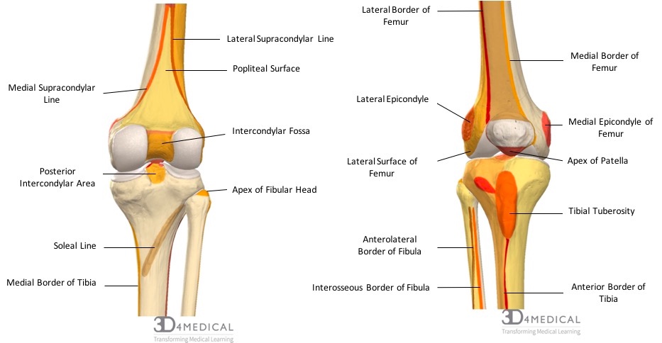
The apex of the fibula is the narrow, pointed end of the bone located on the lateral side of the lower leg. It is situated just below the knee joint and is an important part of the ankle and foot.
The fibula is a long, thin bone that runs parallel to the tibia, the larger bone in the lower leg. It is located on the outside of the tibia and is responsible for providing stability to the ankle and foot. The fibula also plays a role in the movement of the ankle and foot, particularly when walking or running.
The apex of the fibula is the topmost point of the bone and is located just below the knee joint. It is a small, narrow structure that is easy to overlook, but it is an important part of the anatomy of the lower leg. It is connected to the tibia by a series of ligaments and muscles, which help to support and stabilize the ankle and foot.
The apex of the fibula is also an important attachment point for several muscles and tendons in the lower leg. These muscles and tendons work together to control the movement of the ankle and foot, allowing us to walk, run, and perform other activities that involve the lower legs.
In addition to its role in movement and stability, the apex of the fibula also plays a role in protecting the lower leg from injury. The bone is surrounded by a thick layer of connective tissue, which helps to absorb shock and protect the leg from impact.
Overall, the apex of the fibula is an important part of the lower leg that plays a vital role in movement, stability, and protection. Without this small but important structure, our ability to walk and perform other activities that involve the lower legs would be greatly impaired.
Fibular Head Pain? Here's What to Do!
The fibula and the tibia join together at the knee and ankle joints. When these ligaments become too loose this can cause the fibula to become unstable and fibular head pain. How do you know what side your fibula is? These fibulae Diese abgerundeten Bogenfibeln wurden erstmals im 12. An important question that pops up on a lot of anatomy tests is with what bony structure does the head of the fibula articulate? The superficial fibular peroneal nerve provides the motor supply to the fibularis longus and sensory supply to the skin of the lower anterolateral aspect of the leg. Soft tissue refers to muscles, tendons, ligaments, and other types of tissue that surround and connect your bones. The intramedullary nail is screwed to the bone at both ends. If possible, your healthcare provider can realign your broken bones without surgery.
Fibula Anatomy : Bony Landmarks & Muscle Attachment » How To Relief
No evidence of an avulsed bone fragment originating from the site of attachment of the lateral collateral ligament or the tendon of the biceps femoris muscle was revealed on MR imaging or at explorative surgery. Surgical Repair of the Distal Fibula Complete fractures and orthopedic injuries to the distal fibula, including those of the tibiofibular syndesmosis, often require surgical repair and fixation with screws and plates. The fibula is a little bone that can cause quite a bit of trouble. . The attachment leaves a gap at the upper end of the passage of the anterior tibial vessels. This type of injury is known as a stress fracture.
The most common clinical complaints were pain in the posterolateral aspect of the knee and a sensation of the knee giving way during walking or running. It may also have reduced the sensitivity for detecting acute ligamentous injury. Immediately below the head, the fibula constricts and the part is referred to as neck of the fibula. The diagnosis is usually established by physical examination and confirmed by electrodiagnostic tools. Jaypee Brothers Medical Publishers. Tendons are thick pieces of connective tissue that connect muscle to bone.
The fibula is a long bone, meaning that it is longer than it is wide. The table below summarizes the muscles that originate from, and insert on the fibula. . On the other hand, the surfaces are the flattened areas that exist between the borders. It is important to note that the common fibular nerve gives off a small sensory branch, the lateral sural cutaneous nerve, which provides sensation inferolaterally to the knee. Radiographically, the bony fragment was horizontally oriented and similar in size in most patients, ranging from 8 to 10 mm in length and from 2 to 5 mm in width.
Common fibular (peroneal) nerve: origin, course, function
Joints are typically hypermobile with excessive joint range of motion because of a defect in collagen formation. View larger version 189K Fig. Note avulsion fracture of posterior tibial plateau at attachment of posterior cruciate ligament short arrow, D in coronal STIR image. Fallen fibula , Mit einem gebrochenen Schienbein in den Fluss gefallen, erklärte, warum sie es nie zurück zum Ufer geschafft hat. This classification was later rearranged and became more widely used in 1972 thanks to Bernhard Georg Weber a Swiss orthopedic surgeon.
Apex of head of fibula: Definition with Apex of head of fibula Pictures and Photos
In addition, if the problem is an irritated spinal nerve in the low back, then an epidural injection can be used to treat that problem 14. EDS has many different signs and symptoms which can vary significantly depending upon the type of EDS and its severity. Despite fusion of the bones, side-to-side motion is present in FH due to an abnormal, ball-and-socket shaping of the ankle joint. Avulsion fractures are strongly associated with disruption of the posterior cruciate ligament. The size of the fragment ranged from 8 to 10 mm in length and from 2 to 5 mm in width.
Fibula: Anatomy, bone landmarks and clinical aspects
The fibulas are Die Schien- und Wadenbeine sind gebrochen. The arcuate ligament inserts in the fibular styloid process deep relative to the fabellofibular ligament. Similarly, superficial and deep fibular nerves, which innervate the muscles attached to the fibula, also innervate the fibular Clinical points Fractures of the fibula are most likely related to traumatic injuries. The deep fibular peroneal nerve, on the other hand, mainly supplies the muscles of the anterior compartment of the leg and the dorsum of the This article will discuss the Key facts about the common fibular peroneal nerve Origin Sciatic nerve; root value L4-S2 Branches Articular branches, lateral sural cutaneous nerve, sural communicating branch, superficial fibular nerve, deep fibular nerve Supply Motor: Anterior leg muscles tibialis anterior, extensor hallucis longus, extensor digitorum longus ; lateral leg muscles fibularis longus, fibularis brevis ; dorsal foot muscles extensor digitorum brevis, extensor hallucis brevis Sensory: Skin of anterolateral leg and dorsum of foot, skin of web space between great and 2nd toes Synonyms: Posterior root of sciatic nerve, Common peroneal division of sciatic nerve , The common fibular peroneal nerve root value L4-S2 is the smaller of two terminal branches of the sciatic nerve, the other being the The nerve arises from the sciatic nerve at the distal third of the anterior compartment of the leg. Symptoms and signs associated with benign and malignant proximal fibular tumors: a clinicopathological analysis of 52 cases.
Sports Med Arthrosc Rev. Can I drive with a fractured fibula? This delay reduced the incidence of bone marrow edema, which was identified in the fibula in only three of 13 patients. None of the patients had a tear of the tendon of the biceps femoris muscle. The lateral compartment of the leg is formed by the muscles responsible for the eversion of the foot and includes the fibularis longus and fibularis brevis muscles. Why do my legs hurt when I break it years? Watch my video below to understand that better: Ehlers-Danlos Syndrome EDS Disorders that affect and weaken the connective tissues such as tendons and ligaments. Importantly, the The function of the proximal end of the fibula is to provide points of attachment for minor supporting ligaments of the fibular collateral ligament that arises from the fibular apex and is surrounded by the tendon of Shaft The majority of the fibula is made up by its shaft.