Bright field microscope parts. What are the parts of the brightfield microscope? PreLab 3.8 2023-01-01
Bright field microscope parts Rating:
7,7/10
1051
reviews
A bright field microscope is a type of optical microscope that uses light to illuminate the specimen being viewed. It is one of the most common types of microscopes used in laboratories and classrooms, and is often the first type of microscope that students encounter.
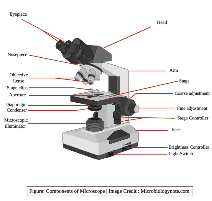
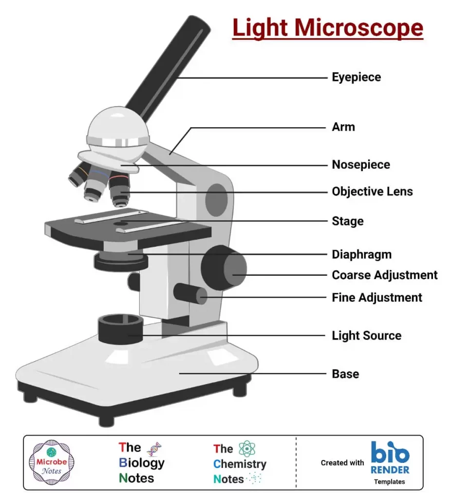
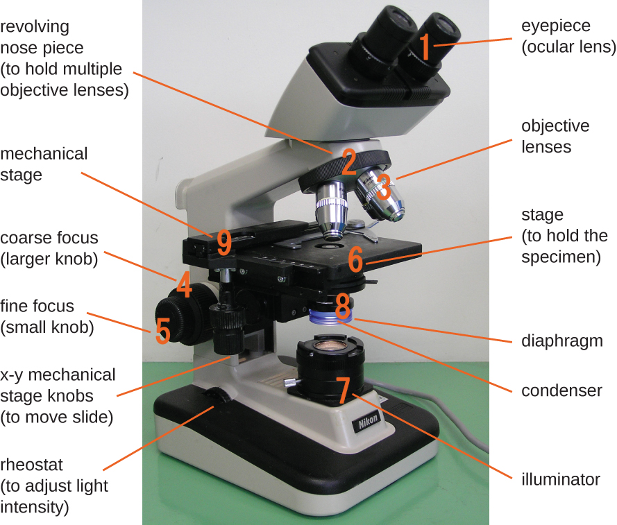
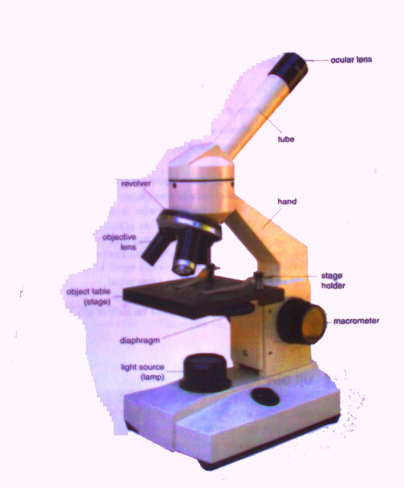
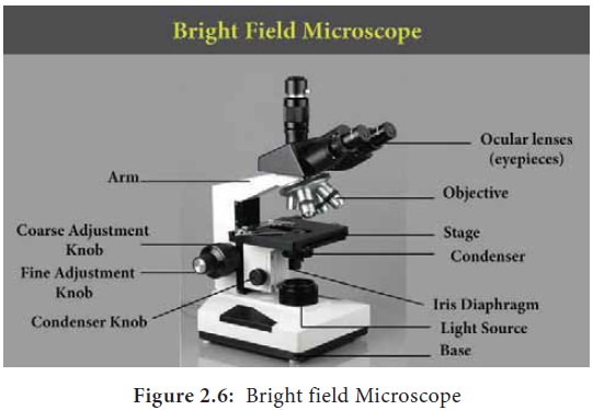
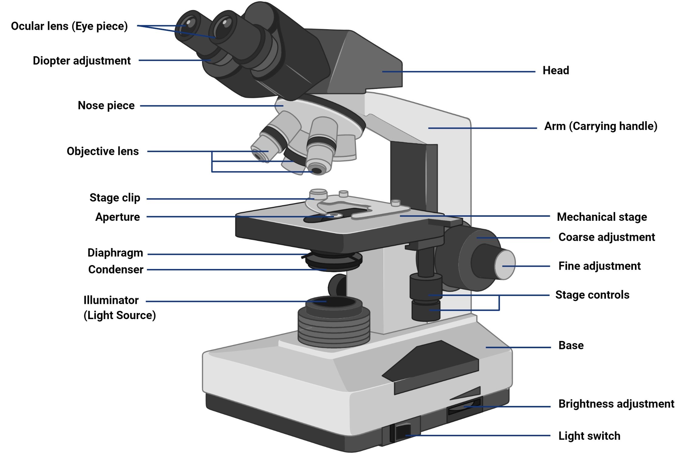
The main parts of a bright field microscope include the following:
Eyepieces: These are the lenses that the user looks through to view the specimen. Most bright field microscopes have two eyepieces, which can be adjusted for focus and magnification.
Objective lenses: These are the lenses that are closest to the specimen and are used to focus the light onto the specimen. Bright field microscopes typically have multiple objective lenses of different magnifications, which can be selected by the user.
Stage: This is the platform on which the specimen is placed. It is usually made of glass and is transparent, allowing light to pass through and illuminate the specimen. The stage is often adjustable, allowing the user to move the specimen in different directions for better viewing.
Condenser: This is a lens system located beneath the stage that concentrates the light onto the specimen. The condenser can be adjusted to control the intensity and focus of the light on the specimen.
Light source: This is the source of light that is used to illuminate the specimen. Most bright field microscopes use a bulb or LED light source, which can be adjusted for intensity and color temperature.
Diaphragm: This is a device that controls the amount of light that passes through the condenser and onto the specimen. It is typically adjusted using a lever or knob on the microscope.
Body: This is the main structure of the microscope, which houses all of the other parts and provides support for the user. The body of a bright field microscope is usually made of metal or plastic, and may be mounted on a stand or tripod.
In addition to these main parts, bright field microscopes may also have other features such as a mechanical stage, which allows the user to move the specimen with precise control, and a built-in camera or video system, which allows the user to capture images or video of the specimen being viewed.
Overall, the bright field microscope is a powerful and versatile tool for examining a wide range of specimens at high magnifications. Its main parts work together to produce bright and clear images, making it an essential tool for many scientific and educational applications.
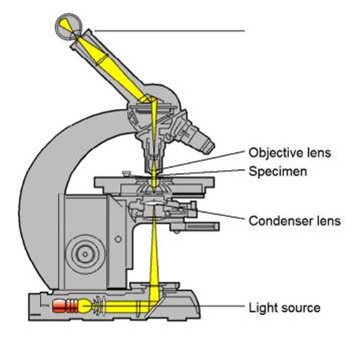
The technique can be used either online or off-line from video recording. It is a type of light microscopy, where a path of light is very simple, which requires a light source like a halogen lamp , condenser lens, objective lens and ocular lens. Biologists estimate that 380 trillion viruses are living on and inside your body right now—10 times the number of bacteria. It is a tool wonderful in the field of research as it allows the observation of cellular tissues or cells alone. Coaxial Focus : A focusing system with both the coarse and fine focusing knobs mounted on the same axis.
Parts of a microscope with functions and labeled diagram
It is useful in various fields of sciences such as physical and biological science, nanotechnology, metallurgy, and forensic analysis. Total magnification of a microscope is determined by multiplying the magnification capability of the eyepiece lens by that of the objective lens. Body tube Head : The body tube connects the eyepiece to the objective lenses. The condenser is a lens system located below the stage that focuses the light onto the specimen. That means light from the object you are viewing passes through two lenses before it reaches your eye.
It comes with a hole in the center that enables the light to pass from below. To identify the bacteria, one can perform gram staining. Binocular Microscope : A microscope with a head that has two eyepiece lenses. Overall, microscopes play a vital role in many scientific and medical fields, allowing us to study and understand the world around us at a level of detail that would be otherwise impossible. They play a major role in ensuring clear sharp images are produced with a high magnification of 400X and above.
What is Bright Field Microscopy? Definition, Steps & Working
Serologic studies lack sensitivity and specificity. And it is this very simplicity what makes it one of the most popular in use. Scanning probe microscopes and electron microscopes are commonly used to study the properties of nanomaterials. Examples are given below: Objective magnification Eyepiece magnification Total magnification 10X 10X 100X 40X 10X 400X 100X 10X 1000X Applications of Microscopes Microscopes are used in a wide range of scientific and medical fields, including biology, medicine, materials science, and nanotechnology. Paul C Johnson, in Microcirculation, 2008 I.
Bright field Microscope: Facts and FAQs » Microscope Club
Parasitology Research 106: 213—219. Microscope Definition Microscopes are instruments that are used in science laboratories to visualize very minute objects such as cells, and microorganisms, giving a contrasting image that is magnified. The transformer regulates the voltage that will reach the lamp -Mechanic system The tube It is a hollow black cylinder through which the light beams travel until they reach the eyepiece. For example, compound light microscopes have compound lenses- an objective lens and an ocular lens. Magnification : The essence of a microscope is its ability to magnify a specimen. The field diaphragm controls how much light enters the substage condenser and, consequently, the rest of the microscope. What is a bright field image? A polarized microscope is a common tool in pathology laboratories.
Most of the time, the body can move up and down and around the pole. The fine adjustment knob is used to bring the image into sharp focus. The simple polarized microscope appears to be the best tool for MSU crystal detection, showing crystals that are not apparent on bright-field observation. Reticle : A small glass circle etched by laser with fine measurements and placed within the eyepiece in order to enable actual measurements of the specimen to be taken. It is capable of displaying both stained and unstained images.
The two sporocysts can be observed Figure 2 a ; however, the oocyst wall is barely visible. The coarse and fine knobs also sharpen the image. Bright-field microscope is a compound light microscope, which illuminates the background against a stained specimen. Most of the time, the larger knob on the outside is the coarse focus, and vice versa. Principle of Brightfield Microscope The principle of a brightfield microscope is based on the use of visible light and an objective lens to magnify and illuminate a sample. T-Mount: A standard adapter for mounting 35mm cameras to microscopes.
Spring loaded objective lenses will retract if the objective lens hits a slide, preventing damage to both the lens and the slide. In the bright field image the unscattered transmitted electron beam is selected with the aperture, and the scattered electrons are blocked. The eyepiece is typically equipped with a focusing mechanism that allows the user to fine-tune the focus of the image. The placement of the eyepiece is such that its eye upper lens further magnifies the real image projected by the objective. Some CPP crystals showing little brilliance under polarized light show enough color change under the compensated polarized microscope to be identifiable. They also hold the microscope base which is the stand of the microscope.
What are the parts of a brightfield light microscope?
It is useful to observe these changes as a learning experience regarding the possible shapes presented by CPP crystals under the microscope, since these orientation-dependent, less characteristic shapes are usual. It consists of a series of overlapping metal or glass blades that can be adjusted to allow more or less light to pass through. The eyepiece is typically located at the top of the microscope, and the objective lens is located at the bottom, near the object or sample being viewed. The specimens used are prepared initially by staining to introduce color for easy contracting characterization. There is a coarse adjustment knob on the arm of the microscope that is used to move the nosepiece or the stage to focus on the image.