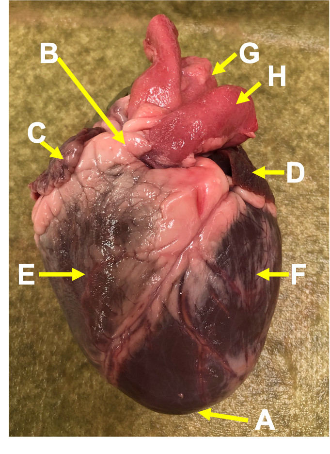
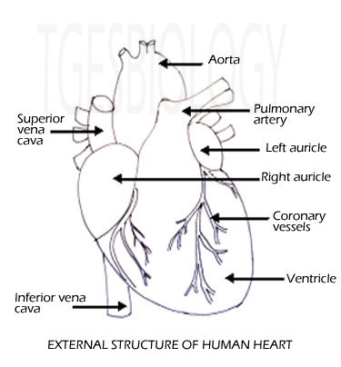
External structure of human heart. Internal and external structure of the Human Heart 2022-12-18

External structure of human heart Rating: 5,8/10 1543 reviews

The human heart is a complex and vital organ that is responsible for pumping blood throughout the body. It is located in the thoracic cavity, between the lungs, and is protected by the ribcage. The heart is divided into four chambers: the left and right atria, and the left and right ventricles. The atria are the upper chambers of the heart, while the ventricles are the lower chambers.

The heart has a unique external structure that helps it to function effectively. The outer layer of the heart is known as the epicardium, which is a thin layer of tissue that covers the surface of the heart. The middle layer of the heart is known as the myocardium, which is a thick layer of muscle tissue that is responsible for contracting and relaxing to pump blood through the heart. The inner layer of the heart is known as the endocardium, which is a thin layer of tissue that lines the inside of the heart and helps to prevent damage to the heart muscle.

The heart is also surrounded by a network of blood vessels, including the aorta, the vena cava, and the pulmonary artery and vein. These blood vessels transport oxygenated and deoxygenated blood to and from the heart, respectively.

The heart has a unique system of electrical conduction that helps to coordinate the contraction of the heart muscle. This system includes the sinoatrial (SA) node, the atrioventricular (AV) node, and the Purkinje fibers. The SA node is located in the right atrium and acts as the heart's natural pacemaker, sending electrical impulses that stimulate the contraction of the heart muscle. The AV node is located in the interatrial septum and helps to coordinate the contraction of the atria and ventricles. The Purkinje fibers are located in the ventricles and help to transmit the electrical impulses throughout the heart muscle.

In summary, the human heart has a complex external structure that is essential for its function. It is made up of three layers of tissue, is surrounded by a network of blood vessels, and has a unique system of electrical conduction. All of these components work together to ensure that the heart is able to pump blood efficiently and effectively throughout the body.

Internal and external structure of the Human Heart

external structure of human heart

If the parents have genetic diseases, then their spouse may have the disease as well. The pulmonary artery opens from the right ventricle that carries the blood to the lungs and the aorta arises from the left ventricle carrying the oxygenated blood throughout the body. The myocardium layer gets stretched or thickened because of which the blood is not pumped properly. The heart is enclosed in a pericardial sac that is lined with the parietal layers of a serous membrane. Internal Structure of Heart The internal structure of the heart is rather intricate with several chambers and valves that control the flow of blood.

Next

Human Heart Anatomy

external structure of human heart

. Node along interventricular septum at the top. What is the interior of heart? It is normal during sleep but can be induced by certain drug ingestion and a low body temperature. Right atrio-ventricular valve Tricuspid valve -Located at the opening between right auricle and right ventricle. Blood from the head, Neck and arms is returned to the right atrium by superior vena cava. ADVERTISEMENTS: The human heart is pinkish about the size of a fist and weighs approx.

Next

Anatomy of a Human Heart

external structure of human heart

The heart must generate enough force that it creates more pressure within the ventricle so that blood can move forward to areas of lower pressure i. Blood flows under little pressure in veins. The heart wall consists of three layers: the endocardium, myocardium and epicardium. So any Problem on this Website Contacts Me. Aorta: It arises from left ventricles and carries oxygenated blood to supply it to all body parts. Now from left ventricle oxygenated blood is pumped into aorta through aortic semilunar valve to supply it to all body parts. The two thick walled lower chamber right and left are called ventricles.

Next

The human heart (External and internal structure)

external structure of human heart

The right atrium is located on the superior right side of the heart and is separated from the left atrium by the thin layer called the inter-atrial septum. Thus, The heart consists of four chambers, The right and left atria and the right and left ventricles. The pulmonary trunk divides into two pulmonary arteries one to each lung. Definition of Heart: The heart is the essential organ in the cardiovascular system that is used for the pumping of the oxygenated and deoxygenated blood throughout the body. At the place of crossing the pulmonary aorta and the caroticosystemic aorta are connected by muscular band known as ligamentum arteriosum which during embryonic stages was represented as a single artery known as ductus arteriosum or duct of botalli. The myocardium is the middle layer of the heart.

Next

Heart

external structure of human heart

Artery has thick, muscular and elastic walls. What is the anatomy of the heart? Furthermore, the atria are separated from the ventricles by the atrioventricular septa. It consists of the two mitral leaflets, anterior and posterior sited within the mitral annulus. The atria receive blood and the ventricles distribute it. The walls consist of fewer muscles compared to the left portion, and the size difference is based on their functions. From the aorta blood is distributed to all parts of the body except lungs. Papillary muscles act as anchors for chordae tendinae.

Next

Heart

external structure of human heart

So that this information all subjects of Civil Engineering Students. The left ventricle is the largest of all the chambers and forms… What is the cardiac structure? The heart consists of four chambers, two on the upper side called atria and two on the lower side called ventricles. Furthermore, it prevents the blood from sticking to the inner walls, thereby preventing potentially fatal blood clots. Briefly explain the structure of the human heart. Â There are two sets of valves: atrioventricular and semilunar. A network of muscular ridges called musculi pectinati or trabeculi pectinati present internally in the region of the auricular appendages and give comb like appearance.

Next

Anatomy of the human heart

external structure of human heart

The left atrium receives oxygen-rich blood from the lungs and pumps it to the left ventricle. Human blood circulatory system The human blood circulatory system includes pulmonary circulation, Which circulates blood through the lungs and the systemic circulation, Which serves the needs of the body tissue. Semilunar valves: The aortic valve is present between left ventricle and aorta and Pulmonary valve is present between right ventricle and pulmonary artery. The valve between the right ventricle and right atrium is the tricuspid valve, and the one which is found between the left ventricle and left atrium is known as the mitral valve. Right auricle receives deoxygenated blood from the body parts by anterior and posterior vena cava. The coronary arteries supply oxygenated blood to the heart, and the cardiac veins drain the blood once it has been deoxygenated by the tissues of the heart.


Next

Human Heart

external structure of human heart

This is done by the two coronary arteries: left and right. The heart has four chambers: two atria and two ventricles. The arteries are located in the subepicardium, so they are easily visible in the human eye, but in some cases, especially at their initial course, may be sited within the myocardium. Carotid arteries : Supply blood to the head and neck. The alternate contraction and relaxation dilation continues regularly.

Next

What is internal and external structure of heart?

external structure of human heart

It is about the size of a man's fist. The left ventricle has chordae tendinae and papillary muscles which prevent tricuspid and bicuspid valves from being pushed into auricles at the time of ventricular contraction. The main cause of the pericardial disease is the inflammation of the pericardium, infection with a virus, rheumatoid arthritis, or injury of the pericardium. Weight: In males it weighs: 280-340 gm and in females : 230-280 gm Describe the external features of Heart. Pulmonary Circulation The pulmonary circulation consists of pulmonary trunk, Pulmonary arteries and pulmonary veins. From the right ventricle, the pulmonary aorta arises whereas from the left ventricle systemic aorta arises.

Next