Eye ball layers. eyeball 2022-12-17

Eye ball layers Rating: 9,7/10 889 reviews

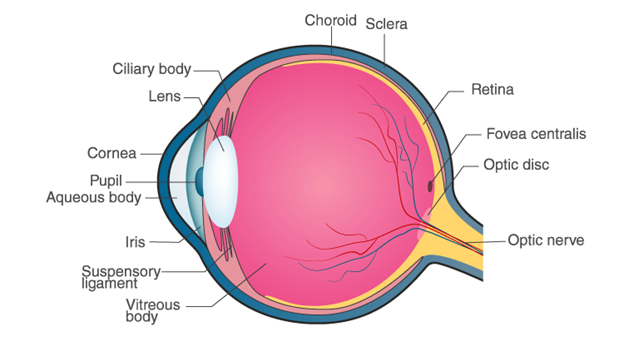
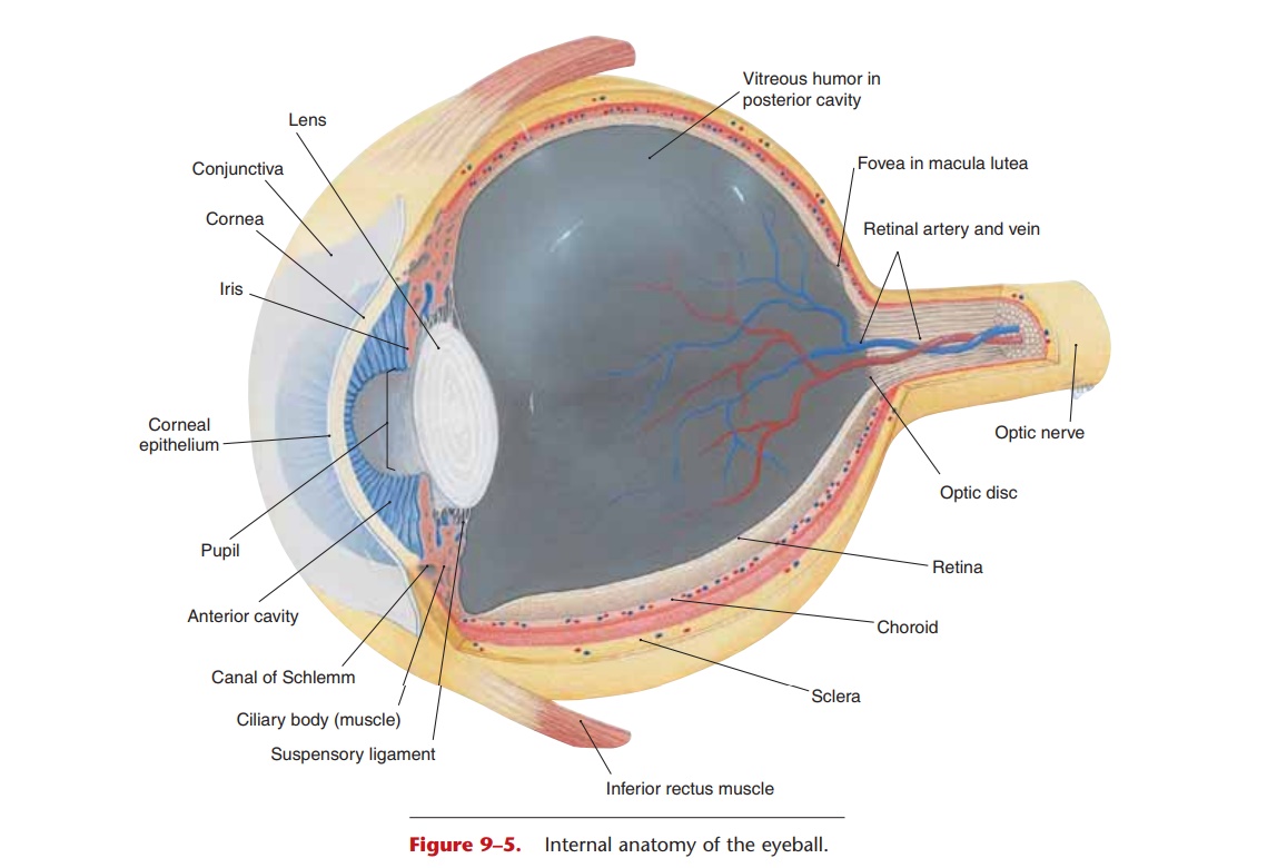
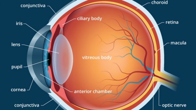
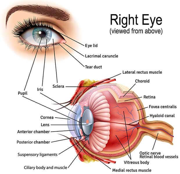
The eye is a complex and fascinating organ that plays a vital role in our ability to see and interpret the world around us. It is made up of several layers, each with its own unique structure and function. Understanding these layers can help us appreciate the intricate design of the eye and the important role it plays in our lives.

The outermost layer of the eye is the sclera, which is a tough and white outer layer that forms the bulk of the eye's structure. It is made up of dense connective tissue and helps to protect the eye from injury. The sclera is also responsible for maintaining the shape of the eye and helping it to stay in place.

Beneath the sclera is the choroid, a layer of blood vessels that helps to nourish the eye. The choroid also contains the iris, a ring-shaped structure that controls the size of the pupil and determines the amount of light that enters the eye. The iris is responsible for the color of the eye and is composed of pigmented cells that give it its characteristic hue.

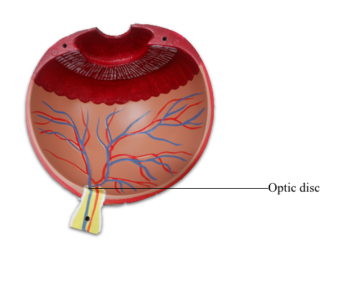
The next layer is the retina, which is a thin layer of light-sensitive cells that line the back of the eye. The retina is responsible for converting light into electrical signals that are sent to the brain via the optic nerve. The retina contains two types of cells called rods and cones, which are responsible for detecting light and color, respectively.

Beneath the retina is the choroid, which is a layer of blood vessels that helps to nourish the eye. The choroid also contains the iris, a ring-shaped structure that controls the size of the pupil and determines the amount of light that enters the eye. The iris is responsible for the color of the eye and is composed of pigmented cells that give it its characteristic hue.

Finally, at the very center of the eye is the lens, which is a clear, flexible structure that helps to focus light onto the retina. The lens is able to change its shape in order to adjust the focus of the eye, allowing us to see objects both near and far.

In summary, the eye is made up of several layers, each with its own unique structure and function. The sclera is the outermost layer and helps to protect the eye and maintain its shape. The choroid contains the iris and helps to nourish the eye. The retina is a thin layer of light-sensitive cells that converts light into electrical signals, and the lens is a clear, flexible structure that helps to focus light onto the retina. Together, these layers work together to allow us to see and interpret the world around us.

What are the 3 layers that make up the wall of the eyeball?

eye ball layers

These are the light-sensitive retinal cells that provide high visual acuity. Kenhub does not provide medical advice. This muscle is responsible for depressing the adducted eye. It rotates the eye outwards front to back and upwards. Retinoblastoma Retinoblastoma is an eye cancer. The shape of the lens is changed by the ciliary muscle, which enables the eye to focus light from objects at varying distances from the eye.


Next

Eyes: Structure, function, and disease

eye ball layers

Similarly, obstruction of the Glaucoma Glaucoma is a condition of increased pressure within the eyeball, which results in gradual loss of sight. Presbyopia This condition occurs due to age and causes a reduction in the focusing power of the lens. Light-sensitive tissues These include the retina and the optic nerve. The retina is the light-sensitive membrane covering the back of the eye. The clear bulging surface of the eye. Edinburgh: Elsevier Churchill Livingstone. Structure of the Eye The cornea is the transparent but alive, curved structure at the front of the eye, with embedded nerve endings for pain, touch and thermal sensation.

Next

Eye Anatomy: Parts of the Eye and How We See

eye ball layers

Its core is looser and features a narrow and somewhat oblique channel that extends from the optic disc to the posterior pole of the lens. Here, the inner retina is firmly attached to the outer RPE. This increases the scope of our view. Skin that covers the upper part of the eyeball, including the cornea, when closed. The medial rectus muscle, for example, pulls the eyeball medially, as if to look at the nose. Aqueous humor is formed by capillaries in the ciliary body, flows anteriorly through the pupil, and is reab-sorbed by the canal of Schlemm small veins also called the scleral venous sinus at the junction of the iris and cornea.

Next

Eyeball: Layers and Cavities of the Eyeball

eye ball layers

Vitreous body Synonyms: Vitreous humour, vitreous humor The vitreous body is the largest structure of the eyeball, occupying the four-fifths of the entire eye. They are : The fibrous coat or sclera protects the intraocular structures. Although the episcleral potential space exists between the fascia and the eyeball, there is actually very little movement between the eye and the sheath, meaning that the fascia and the eye move together within the orbital fat. The iris contains two dilator pupillae muscle. This chamber is a fluid-filled area between the cornea and iris. It contains blood vessels and gives the inner eye a dark color.

Next

Eyeball Anatomy

eye ball layers

The retinalines the posterior two-thirds of the eyeball and contains the visual receptors, the rods and cones Fig. Signs of presbyopia typically begin around the age of 40 and gradually progress until age 65 or 70, when presbyopia plateaus. They also give off smaller branches that supply the circle of Zinn-Haller. Blood supply and innervation Synonyms: Major circulus arteriosus of iris The root of the iris contains a circular anastomotic arterial network called the major arterial circle, formed by anterior and posterior ciliary arteries. This allows the lens to increase its anterior curvature which results in the increase in refractive power. Conjunctiva The Conjunctivitis, often known as pink eye, occurs when this thin membrane becomes inflamed or swollen.


Next

Anatomy of the Eye

eye ball layers

These remarkable features of our eye are enabled by the complex structure of the eyeball. The eye is unable to abduct beyond the midline. Refractive media of the eyeball The refractive media of the eye are the structures that help in focusing the ray of light onto the retina where it can be detected by the photoreceptors. Diabetic Retinopathy This is a common disease among people with diabetes. All of these developments make it difficult for the eyes to focus on nearby objects.

Next

Eye Anatomy: 16 Parts of the Eye & Their Functions

eye ball layers

Refracting tissues Refracting tissues focus incoming light onto light-sensitive tissues to give a clear, sharp image. Composed of retinal nerve fibers that exit the eyeball through the optic disc, traverse the orbit, and pass through the optic foramen into the cranial cavity, where they meet fibers from the other optic nerve at the optic chiasm. Â The venous drainage mirrors the arterial supply; small veins from the pupillary margin form the minor venous circle, from which the larger veins convey the blood into the vorticose veins. Retinal hemorrhages- Diabetes and hypertension cause damage to the retinal arteries. Do you want to learn the blood vessels of the eye using videos, illustrations and quizzes? Optic Disc The optic disc is the place at which the axons of retinal ganglion cells join together and mark the beginning of the optic nerve second cranial nerve.

Next

What are the 3 layers of the eye and their functions?

eye ball layers

Within the neural tunic is the retina, with three layers of cells and two synaptic layers in between. This allows the eye to take in as much light as possible. Fovea Centralis The macula. It arises from the front of the socket near the nose and travels inwards, attaching to the bottom surface of the eyeball. Â The preservation of corneal transparency is very important for maintaining its proper function. The clear part of the eye that helps with focus. In normal conditions, the layers adhere to each other and this space is empty.

Next