Function of a reflex arc. Primary Function Of A Reflex Arc 2022-12-15
Function of a reflex arc Rating:
6,2/10
1525
reviews
A reflex arc is a neural pathway that allows an organism to respond to a stimulus in a rapid, automatic manner. It is a key component of the body's reflexes, which are involuntary responses to stimuli that occur without conscious thought or effort.
The function of a reflex arc is to provide a quick and efficient way for the body to respond to stimuli that may be harmful or dangerous. For example, if a person touches a hot stove, the reflex arc allows their hand to quickly and automatically withdraw from the heat before they are burned. Similarly, if an animal is confronted by a predator, the reflex arc allows it to quickly escape or defend itself without having to consciously think about the action.
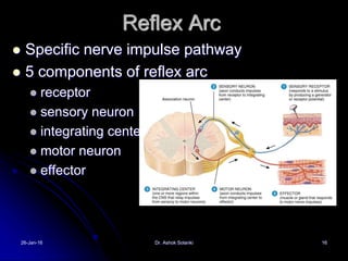
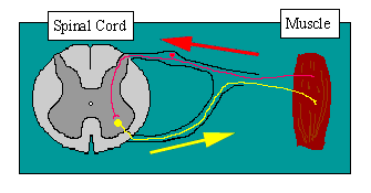
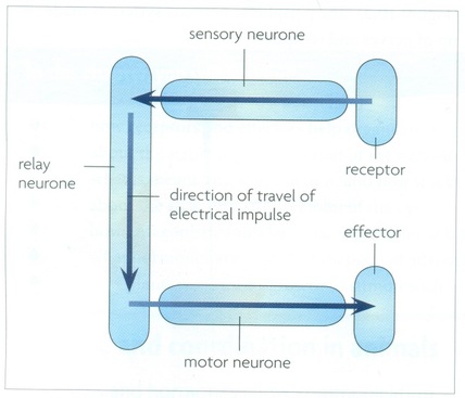
The reflex arc consists of five main components: the sensory neuron, the sensory receptor, the integration center, the motor neuron, and the effector. The sensory neuron receives the stimulus and transmits it to the integration center, which is typically located in the spinal cord. The integration center receives the stimulus and processes it, determining the appropriate response. If the integration center determines that a reflex response is necessary, it sends a signal to the motor neuron, which carries the signal to the effector (such as a muscle or gland). The effector then responds by contracting or releasing, causing the reflex action to occur.
One of the main advantages of a reflex arc is its speed. Because it bypasses the brain and directly connects the sensory receptors to the effectors, reflexes can occur much faster than voluntary actions. This is important in situations where a quick response is necessary to avoid harm or danger.
In summary, the function of a reflex arc is to provide a quick and automatic response to stimuli that may be harmful or dangerous. It consists of several components that work together to receive, process, and respond to the stimulus, allowing the body to react without conscious thought or effort.
Structure and function of the reflex arc
That light is our stimulus, and it hits photoreceptors in the back of the eye. The path taken by the nerve impulses in a reflex is called a reflex arc. Interneurons primarily serve as a pit stop between sensory neurons and motor neurons. In a reflex arc, the sensory neuron sends a signal to the interneuron and activates it. This is because the knee jerk reflex is a stretch reflex that's designed to test the integrity of your lower spinal cord. Once again, interneurons are necessary for coordinating sensory information in this case, pain and generating the appropriate muscular response. Common pathologies include lumbar spinal stenosis, lumbosacral meningomyelocele, or following abdominoperineal resection or radical hysterectomy.
What is the path of reflex action? The knife was swiftly applied to the tendon right below the knee with enough pressure to illicit a reflex, but not enough pressure to cause pain. Among the most common receptors we can find photoreceptors, the cells in charge of detecting light intensity; thermoreceptors, responsible for detecting heat and temperature changes; or mechanoreceptors, neurons that react to mechanical pressure. Moreover, respiration is an essential and vital process for maintaining the proper functioning of the organ system. During that time, she taught several courses including Human Physiology, Introduction to Neuroscience, and Human Biology of Health and Disease. It is used to assess the background tone sent by the brain to the spinal cord. In this article we explain what a reflex arc is, what its main characteristics are, its structure and its components, the functions they perform, as well as the different types of reflections that exist. How do reflexes work? On examination, the bulbocavernosus and superficial anal reflexes are variably absent, decreased, or present.
What Is a Reflex Arc? What happens during inspiration? Reflex Arc Now, let's look at the parts of a reflex. Inspiration occurs via active contraction of muscles — such as the diaphragm — whereas expiration tends to be passive, unless it is forced. In this article, we will discuss the components of the reflex arc, some examples of reflex actions, their importance, and associated clinical conditions. Since the combined effects may cause a very powerful shortening reaction of the stretched muscle, the integrity of its muscle fibres may be endangered in states of hyper-reflexia. However, sometimes it might have only two neurons i. The nerve impulse generated by the stimulus in the sensory neuron travels to the muscle via the interneuron and motor neuron to carry out the reflex action. For example, the withdrawal reflex nociceptive or flexor withdrawal reflex is a spinal reflex intended to protect the body from damaging stimuli.
This classification system does not often reflect clinical practice and deviations from these descriptions often exist. In a normal adult, neurons react to signals that they receive from hundreds of other neurons. Neurons The sensory neuron attached to the Golgi tendon organ synapses has its cell body in the posterior root ganglion. Let's go through each of these components. Although we think of the brain as being the boss of all of our actions and thoughts, some actions actually take place without the brain's input. A reflex is an involuntary response to an external stimulus. On the other hand, they can also have to do with preventing damage to some of our most important organs.
Unlike most neural pathways, in almost all animal species, these do not pass through the brain. Neural Circuit The reflex arc of this reflex action is simple. Afferent sensory vagus neurons transmit peripheral signals to brainstem nuclei; efferent motor vagus neurons project to the periphery and signal primarily via acetylcholinesterase both at pre- and postganglionic neurons. This abundance of interneurons is due to the complexity of integrating the sensory and motor segments of the nervous system and the diversity of functions that exist in the brain and spinal cord. Although older studies suggested that this finding arose from compression of the third cranial nerve by the herniating temporal lobe, the unilaterally dilated pupil appears to develop before actual movement of the medial temporal structures over the tentorial edge. A reflex action is an involuntary response to a stimulus, mostly a pain stimulus, that takes place instantaneously.
Effector Organ An effector organ is one that acts in response to the stimulus. We're going to dive deeper into how reflexes work, the components of them, and how they protect the body from harm. The information goes to your brain for processing before you actually respond. Knee-jerk Reflex External Pain Reflexes Imagine that you accidentally touch something hot. The stimulus, such as a needle stick, stimulates the pain receptors of the skin, which initiate an impulse in a sensory neuron. The result is a contraction of the quadriceps femoris, which causes the lower leg to kick gently. This characteristic allows reflex actions to occur relatively quickly by activating spinal motor neurons without the delay of routing signals through the brain, although the brain will receive sensory input while the reflex action occurs.
The peripheral process of such neurons is long and make connections with the receptor or have a specialized nerve ending that itself acts as a receptor. The pathways for the stapedius reflex to one side ipsilateral side are shown in Fig. The withdrawal reflex allows us to quickly remove parts of our body from painful stimuli. For example, behavior-dependent increases in blood pressure are enabled and moderated by the baroreflex Janig and Habler, 2003 and failure of this autonomic reflex arc causes loss of consciousness due to cerebral hypoperfusion. The intermediate neurons that connect the afferent with the efferent and with the integrating centers, two of the components of the reflex arcs, are known as interneurons. We use a reflex hammer to tap the patellar tendon, which is made up of the tendon of the quadriceps muscles. When something like this happens, it feels like you simply react to the situation automatically, without thinking.
From here impulses are sent to the organs in this example the arm muscle by the spinal nerves. What is the most basic function of respiration? The receptor is present in the receptor organ. Unilateral loss of pupillary constriction in the comatose patient may rarely indicate subarachnoid hemorrhage from an internal carotid aneurysm that compresses cranial nerve III at the origin of the posterior communicating artery Video 32, Cranial Nerve III Palsy. Let's start by acknowledging that reflexes come in many varieties, some more complex than others. While there are many different types of reflexes, they all follow the same basic pattern of a reflex arc.
These inhibitory interneurons inhibit the alpha motor neurons to cause muscle relaxation. The reflex arc in the case of a withdrawal reflex involves the following. When it is stimulated by a motor neuron, the muscle contracts and the body is flexed, moving it away from the stimulus. What is a reflex arc Class 10? Sensory or afferent neurons Once the receptors have captured the information from the outside, the sensory or afferent neurons are responsible for collecting it and transmitting it to the nerve centers the gray matter of the spinal cord, the place where the information is going to be processed in order to elaborate the answer that best adapts to environmental demands. The activated sensory neuron extends into the spinal cord, sending an electrical signal all the way to another neuron, the interneuron. Neural Circuit When tension in muscle increases, the Golgi tendon organ sends negative feedback to the interneurons. Imagine someone has a hold of your knee and is pulling it into an awkward position.