Gram staining experiment conclusion. Conclusion for gram staining experiment 2023-01-01

Gram staining experiment conclusion Rating: 6,6/10 492 reviews

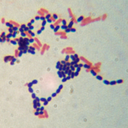
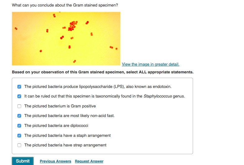
Gram staining is a laboratory technique used to identify and differentiate bacterial species based on their cell wall composition. The procedure involves the application of crystal violet, iodine, and a decolorizing agent to a bacterial sample, which is then observed under a microscope to determine the bacterial species. The Gram stain is named after Danish bacteriologist Hans Christian Gram, who developed the technique in the late 1800s.

The Gram stain is a widely used and reliable method for identifying bacterial species, and it has many practical applications in the fields of medicine, agriculture, and food science. For example, Gram staining can be used to identify the presence of specific pathogenic bacteria in a patient sample, to detect foodborne pathogens in food samples, or to identify the presence of bacteria in soil or water.

One of the key advantages of the Gram stain is that it is a simple, inexpensive, and rapid method for identifying bacterial species. The procedure can be completed in just a few hours, and the results can be observed under a microscope, making it easy to confirm the identity of the bacteria.

Despite its many advantages, the Gram stain is not perfect. It can produce false positive or false negative results, particularly in the case of certain types of bacteria that do not retain the crystal violet-iodine complex. In these cases, other methods such as biochemical testing or DNA sequencing may be needed to accurately identify the bacterial species.

In conclusion, the Gram stain is a valuable and widely used laboratory technique for identifying and differentiating bacterial species. Its simplicity, speed, and low cost make it an important tool for a variety of applications in the fields of medicine, agriculture, and food science. However, it is not foolproof, and other methods may be needed to confirm the identity of certain types of bacteria.

Gram Staining Lab Report Essay Essay

gram staining experiment conclusion

Then, by slow rotation of the loop in the drop, a bacteria suspension is made and it is spread till a smear is obtained. In gram-positive cells, the decolourising agent acts initially as a lipid solvent, dissolving out the little quantity of lipid present in the cell wall, thereby forming minute pores in it. Keep for 24 hours and filter. They are two distinct morphological groups of bacteria. Do some research and describe one other type of acellular organism.

Next

Gram Staining Lab Report

gram staining experiment conclusion

Hypothesis The purposes of this lab is to examine how the Gram Stain techniques …show more content… In the second step we flood the smear with Crystal Violet for 60 seconds. The medical laboratory scientist then puts together a report and sends it to your healthcare provider. How will the gram stain techniques show morphology of a bacteria cell? A medical laboratory scientist processes the Gram stain, which gives relatively quick results, so healthcare providers can know if bacteria are present, and, if so, the general type s. Bacteria are a large group of one-celled organisms. Retrieved October 9, 2017 from iii. Students will review a brief video and slides to introduce the compound microscope. These features make the Gram stain method a more desirable screening procedure for bacterial vaginosis in a clinic population.

Next

Gram stain conclusion Free Essays

gram staining experiment conclusion

Observing a Gram stain in a light microscope The light microscope is arguably the most valuable research tool in the history of biology. Subsequently, it dehydrates the cell wall proteins peptides , which close the pores. Gram Staining proficiency and should be able to: A. What conditions do Gram stains help diagnose? Differentiation of bacteria into gram-positive and gram-negative groups provides the most important clue for proceeding further in proper direction for identification of unknown bacteria. Healthcare providers typically request a Gram stain along with a bacteria culture when they suspect you have a bacterial, or sometimes fungal, infection. It is a technique used to differentiate types of bacteria using certain physical and chemical characteristics of their cell walls. Gram stains generally provide quick results.

Next

Conclusion Staining of bacteria is important to increase the visualization of

gram staining experiment conclusion

Why was the Gram iodine added to the Gram staining procedure? Peptidoglycan is mainly a polysaccharide composed of two subunits called N-acetyl glucosamine and N-acetyl muramic acid. They can live in different places in your body and on your skin. One thing Premium Staining Gram staining Bacteria Bacteria Classification by Gram Staining Bacteria Classification By Gram Staining THE AMERICAN UNIVERSITY IN CAIRO BIOLOGY DEPARTMENT SCIENCE 453 : BIOLOGY FOR ENGINEERS REPORT No. On one of the students slide you could see a lighter purple with white spaces in the walls of the S. These classifications are very important, due to the fact that the topic of this paper, Vibrio Cholerae, the bacterial source of Cholera disease, is known to be a Gram-negative… Double Unknown Lab Report The following tables are each corresponding to either the Gram-positive sample or Gram-negative sample and include the tests utilized along with their results all in the order each test was performed. The primary stain is crystal violet. Students will also view a video about the Gram Stain procedure and then answer questions related to it.

Next

Gram stain blog.sigma-systems.com

gram staining experiment conclusion

Care is taken, so that, water does not fall directly on the smear. In the third step, we gently emulsify the organism in the drop of liquid spreading the mixture over the entire surface of slide to produce an even thin film of bacterial smear. On what part of the cell do the antibiotics usually work? Different types of bacteria are either gram-negative or gram-positive. More understanding the types and morphology of bacteria. Thus, allowing health professionals to prescribe appropriate antibiotic for adequate treatment. The experiment was conducted to further understand the purpose of a Gram stain. It acts as a lipid solvent and as a protein- dehydrating agent.

Next

Gram Staining : Principle, Procedure, Interpretation and Animation

gram staining experiment conclusion

Using either a textbook or a reputable online resource, research some of the typical characteristics of bacteria, and discuss why it might be important for a researcher or a hospital technician to be able to differentiate between Gram-positive and Gram-negative bacteria. For example, a gynecologist may collect a swab for a gonorrhea Gram stain test and a pulmonologist may collect a sample of your spit or mucus from your cough to use for a bacterial pneumonia Gram stain test. Introduction Gram staining is the most important differential staining method used in microbiology. Do a search online 1-2 antibiotics that affect Gram-positive bacteria and list them. Who performs a Gram stain test? SUBJECT MATTER: Biological Techniques Topic: Staining A.

Next

Conclusion gram staining Free Essays

gram staining experiment conclusion

Methods The method or procedure used in this gram stain lab test for Staphylococcus aureus and Escherichia coli is as elaborated below. The final way to categorize a bacterial cell is by its genetic makeup, or more specifically its ribosomal sequences within its RNA. Gram staining is one of the most useful test used in the clinical setting to identify bacterial colonies due to their broad staining spectrum. The fourth step, we allow the smear to air dry completely. Retrieved December 26, 2017 from ii. The Gram stain is a differential stain that allows you to classify bacteria as either gram positive or gram negative. The strength of keratin also makes it resist Premium Gram Staining Micro Biology Lab Gram Staining Lab Report Purpose The purpose of this lab was to experiment with a staining technique called Gram Staining.

Next

Gram Stain: What It Is, Purpose, Procedure & Results

gram staining experiment conclusion

Finally, Gram-positive bacteria have no outer membrane, while gram-negative have an outer membrane. What do you concluded? Why is a counterstain safranin added to the Gram staining procedure? In this lab, both viruses and prions were introduced as acellular organisms. Rezaga Submitted to: Dr. The smear was subsequently covered with the primary stain known as Crystal Violet and left for one minute. .

Next

What is the conclusion of the gram staining technique?

gram staining experiment conclusion

The bacteria that stains purple are gram-positive and the bacteria that stains pink is the gram-negative bacteria. Figure: 1 step 3. The Gram-staining technique uses two types of stains; a primary color and a counterstain. One of the most fundamental differential staining techniques used in the study of bacteriology is gram staining. Gram-positive are single-layered and smooth cell walls, while Gram-negative have a double-layered cell wall, which is wavy. Results are often available within a few days, but some types of bacteria grow slowly, which may take several days or longer.

Next