Lacrimal bone function. Lacrimal bone anatomy, diagram and function 2022-12-13
Lacrimal bone function Rating:
5,2/10
763
reviews
The lacrimal bone, also known as the small, thin bone located in the eye socket, plays an important role in the anatomy of the face and the function of the eye.
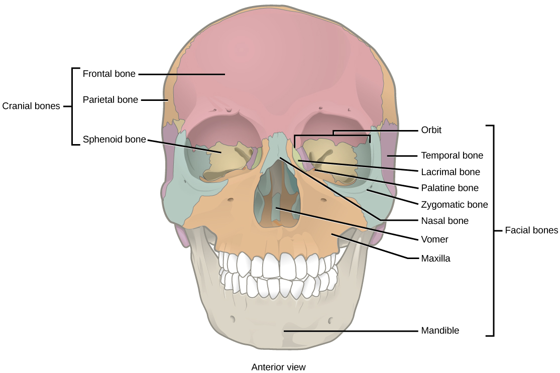
One of the main functions of the lacrimal bone is to provide support and protection to the eye. The lacrimal bone is located in the medial (inner) aspect of the eye socket and helps to form the wall of the orbit, or eye socket. This bony structure helps to protect the eye from injury and also helps to maintain the shape and position of the eye within the orbit.
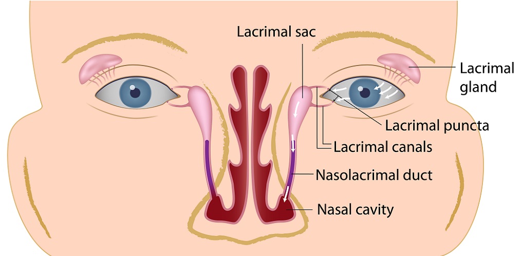
Another important function of the lacrimal bone is in the production and drainage of tears. The lacrimal bone contains a small depression, known as the lacrimal fossa, which houses the lacrimal gland. The lacrimal gland is responsible for the production of tears, which are necessary for maintaining the health of the eye and providing lubrication to the surface of the eye. The lacrimal bone also contains a small opening, called the lacrimal canaliculus, which allows for the drainage of tears from the eye.
In addition to its role in the production and drainage of tears, the lacrimal bone also plays a role in the movement of the eye. The lacrimal bone is connected to several muscles that control the movement of the eye, including the lateral rectus muscle, which allows for movement of the eye outward, and the medial rectus muscle, which allows for movement of the eye inward. These muscles are essential for proper eye movement and coordination.
In conclusion, the lacrimal bone is a small but important bone in the anatomy of the face and the function of the eye. It provides support and protection to the eye, plays a role in the production and drainage of tears, and is connected to several muscles that control eye movement.
What is Lacrimal nerve? Structure, Function, Location and Anatomy
Lacrimal fluid contains proteins, antimicrobial agents, water and electrolytes that ensure adequate lubrication, protection and nutrition of the ocular surface. This is where the two bone surfaces meet. Its posterior region articulates with the ethmoid and meets some of the anterior ethmoidal cells. The nasolacrimal duct tear duct sits between the lacrimal and. The vomer separates the right and left nasal cavities as a small, ridge-like bone. Both bones have two surfaces: the lateral orbital surface and the medial nasal surface.
The Lacrimal Bone: The Anatomy Of Facial Skeleton In Humans
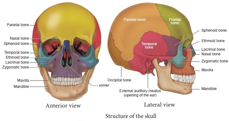
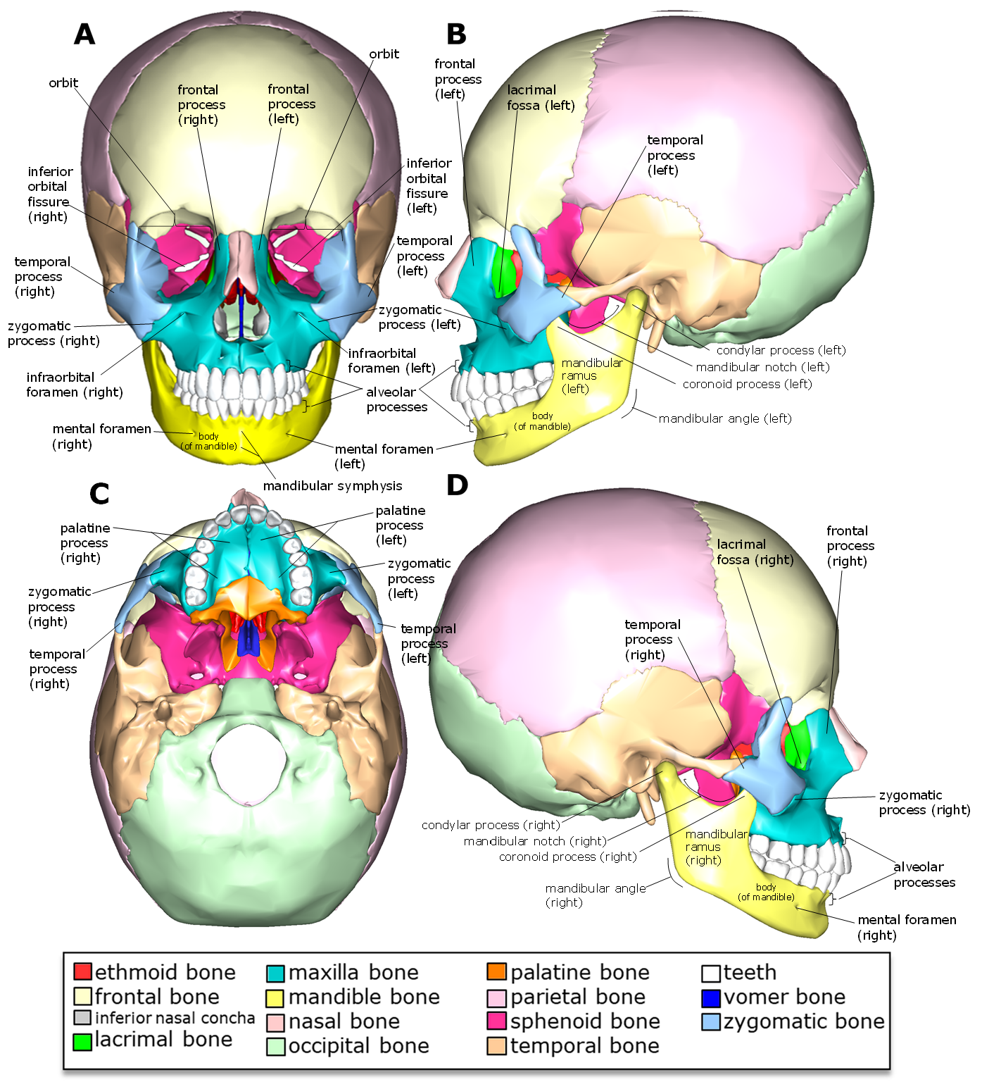
What is a good vitamin for dry eyes? Tears or lacrimal fluid from the lacrimal glands are stored in this sac in cases of excessive lacrimation. Joint The lacrimal bone articulates with four bones: two cranial, frontal and ethmoid, and two facial, maxilla, and inferior nasal concha. Last medically reviewed on January 20, 2018. This can lead to dryness and irritation of the eye, as well as an increased risk of eye infections. To prevent permanent obstruction of the nasolacrimal duct, all lacerations to the tear ducts must be properly repaired. Several bony landmarks of the lacrimal bone function in the process of lacrimation or weeping. The lacrimal bone develops from this single ossification center.
The outer surface forms the anterior part of the inner wall of the orbit, and the inner surface forms part of the outer wall of the nasal cavity middle nasal meatus. Medical science monitor: international medical journal of experimental and clinical research, 20, 628—638. The smooth posterior end of the posterior lacrimal crest forms the medial wall of the eye socket. The posterior lacrimal crest is a small, vertically elevated ridge that forms a groove around the eye the lacrimal sulcus. The Bones of the Face: Our faces are perhaps our most unique feature. Preganglionic parasympathetic fibers from the greater petrosal nerve a branch of the nerve of the pterygoid canal.
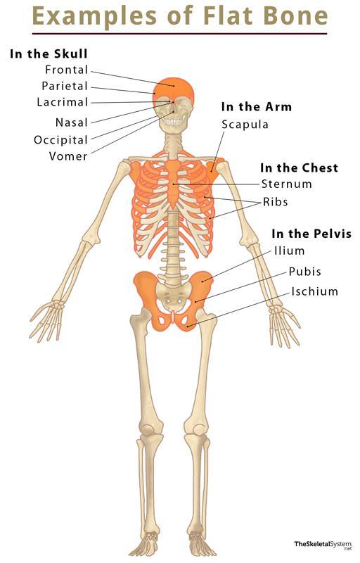
What is the safest eye drops for dry eyes? These joints are found in the frontal, ethmoid, lower nasal concha, and maxilla. Poor secretions should be treated by lid hygiene and massaged with a moist cotton tip in order to remove debris from the eye and increase blood flow so as to open up occluded meibomian glands. This bone is also produced by connective tissue ossification. Borders The lacrimal bone articulates with other skull bones via these four borders. It is caused by meibomian gland dysfunction, which occurs in over 85% of dry eye disease. The bone gets its name from that function; Lacrimal is derived from the Latin word for tears. Facial expression is important for kathakali dancers It is our facial skeleton that supports the skin and mucous membranes.
Does lacrimal gland cause dry eyes? Explained by FAQ Blog
Preocular tear film Lacrimal fluid forms the aqueous component of the tear film, which is a three-layered coating of the ocular surface. Learn faster and improve your understanding with our Lacrimation reflex Stimulation of the cornea and conjunctiva activates a reflex pathway that triggers an increase in tear production from the lacrimal gland. Surgery is avoided when the bones shatter but do not move. Synonyms: Fold of Hasner, Hasner's valve , Anterior to the posterior lacrimal crest is a longitudinally positioned groove called the lacrimal groove or lacrimal sulcus. THE LACRIMAL BONE The Lacrimal Bone: Ever wondered how you tear up and what is involved in the formation of tears? Fractures of this bone often causes obstruction of the nasolacrimal duct. The sympathetic fibers, subsequently, without synapsing in the ganglion, travel along the same path as the parasympathetic fibers supplying the lacrimal gland. The upper part of the lacrimal fossa houses the lacrimal sac, whereas the lower part contains the nasolacrimal duct.
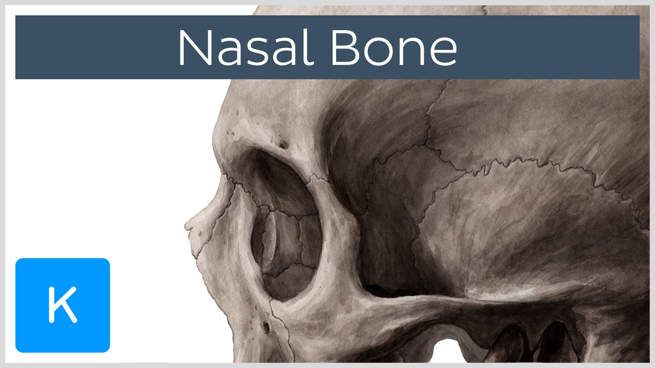
You can see this groove in the picture, which is labeled the lacrimal sac fossa. The sinuses play important roles in our immunity and lung health. At the medial canthal region of the eye, the fluid collects in a triangular space called the lacrimal lake. Edges of the lacrimal bone The lacrimal bone has four boundaries with other craniofacial bones called joints. A 3-D facial bone CT is much preferred by maxillofacial surgeons as this shows distances and distinctly more detail than an x-ray. It is a pair of left and right irregular rectangular thin bones that form the latter half of the fossa for the lacrimal sac at the anterior end of the sidewall of the orbit, which also forms part of the bone wall of the nasolacrimal duct that follows.
Lacrimal Bone: What is it, Location, Function, and More
This circular muscle not only shuts the eyelids, but it also aids tear drainage by pushing tears into the nasolacrimal duct as it contracts. Alternatively, some anatomists do not count the mandible, as this bone is only attached to the skull via ligaments at the temporomandibular joint ; it only directly joins to its paired half through the length of the chin. These are involved in corneal regeneration and maintenance of corneal avascularity and transparency. These ducts open along the lateral aspect of the superior fornix of the conjunctiva. The three types of Le Fort fracture Cranial vs Facial Bones Cranial and facial bones are not the same. They make up the anterior portions of the medial walls of your orbits and span between them. Also with the inferior nasal concha and each other.
The facial bones comprise the anterior and inferior portion of the skull; they extend essentially from the top of your nose to the bottom of your jaw, or mandible, up and down, and from ear to ear side to side, excluding parts of your eye sockets, or orbits. The surgery that opens the duct is known as a dacryocystorhinostomy; a stent is occasionally used to keep the duct open. The orbital surface of the lacrimal bone is divided by a ridge called the posterior lacrimal crest. Ducts from the orbital part of the gland accompany those of the palpebral part by piercing through the levator palpebrae superioris aponeurosis to empty into the conjunctival sac. It's responsible for activities such as biting and mastication, or chewing. Lacrimal Bone The Both paired bones form the border with the maxillary, ethmoid, and frontal bones of the face and skull.
The lacrimal bone has two sides and four edges. Medial surface of the lacrimal bone The medial surface of the lacrimal bone is the part that faces the midline of the body, the posterior surface. Blepharitis, or lid margin inflammation, is both a cause and an effect of meibomian gland dysfunction. The bones in the face are important for the sensory organs located here and for what we look like. The outer surface faces the orbit, and the groove-shaped depression in front of the longitudinal ridge is the part that adds to the composition of the fossa for the lacrimal sac. Which condition is an inflammation of the lacrimal gland? Facial structure is also maintained by articulations and sutures within the zygoma area, with multiple parts of the cranium and parts of the face. The lacrimal sulcus provides space for the soft tissues of the lacrimal sac and the nasolacrimal duct.