Lacrimal sulcus. Nasolacrimal groove. What is this, photo, how to remove, get rid 2022-12-12
Lacrimal sulcus Rating:
9,4/10
703
reviews
The lacrimal sulcus is a small groove or channel located on the surface of the eye, specifically on the upper eyelid. It is located just above the eyelashes and serves as the site where the tears drain into the lacrimal canaliculi, small tubes that lead to the lacrimal sac. The lacrimal sac is a small, pouch-like structure located in the inner corner of the eye socket, where the tears are collected before they are drained into the nasal cavity through the nasolacrimal duct.
The lacrimal sulcus is an important structure in maintaining the overall health and functioning of the eye. Tears are produced by the lacrimal gland, located above the outer corner of the eye, and are essential for maintaining the moisture and lubrication of the surface of the eye. Tears help to wash away any foreign particles, such as dust or debris, that may come into contact with the eye, and also help to keep the eye hydrated and comfortable.
Problems with the lacrimal sulcus can lead to issues with tear drainage and can cause a condition known as epiphora, or excessive tearing. This can be caused by a blockage or obstruction in the lacrimal canaliculi or nasolacrimal duct, or by a deficiency in the production of tears. Epiphora can cause discomfort and irritation, and if left untreated, can lead to further problems such as infection or damage to the eye.
Treatment for problems with the lacrimal sulcus may involve the use of medications or the use of tear substitutes to help maintain moisture on the eye surface. In more severe cases, surgery may be necessary to remove any blockages or to repair any damage to the lacrimal canaliculi or nasolacrimal duct.
Overall, the lacrimal sulcus is an important structure that plays a vital role in maintaining the health and comfort of the eye. Proper care and attention to any issues with this area can help to ensure that the eye remains healthy and functioning properly.
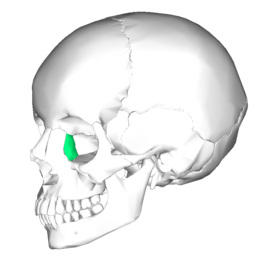
Lacrimal Bone Anatomy, Diagram & Function
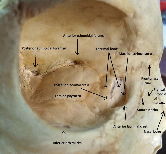
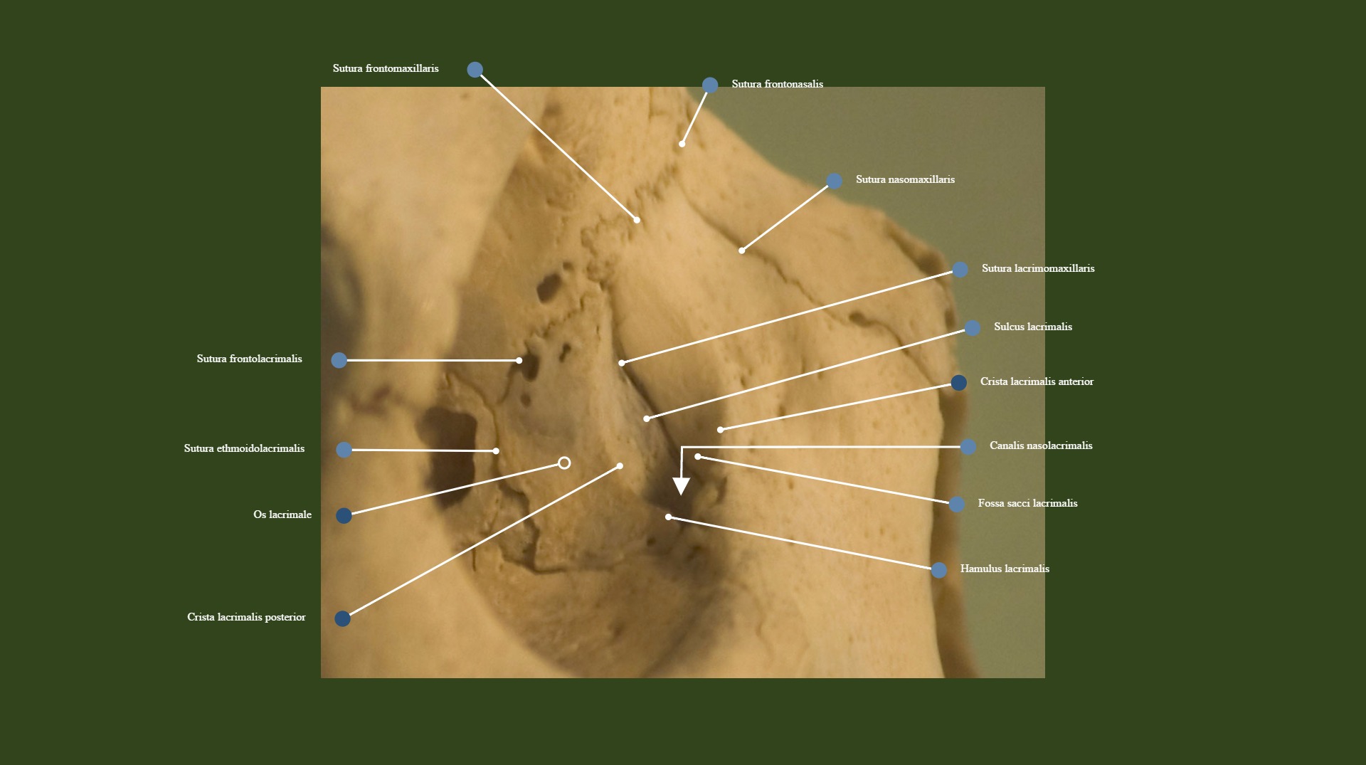
However, the crease can form at a younger age. The anterior crest of the labelled lacrimal bone connects into the maxillary frontal process, and the apex of the bone fits into the frontal bone of the forehead. References Ducker L, Rivera RY. The lacrimal bone is perhaps the most fragile bone in the face and one of the smallest bones in the body. The bone fits snuggly into the eye socket. Perform the procedure 7 times.
Grayson, MD, medical director and chief of glaucoma and cataract surgery at Omni Eye Services in New York and New Jersey, and assistant professor of ophthalmology at the New York Ear and Ear Infirmary of Mt. The composition contains hyaluronic acid with coconut water, which refreshes and moisturizes the skin, eliminating dark circles under the eyes and signs of fatigue. But if you have 180 degrees of support missing, even with a capsular tension ring, anything in there could take a dive. The frontal lobe of the brain and the nasal airway are quite near to the lacrimal bones. Osteoporosis can impair an extremely susceptible structure, and there is a relationship between lacrimal bone density and total bone density.
At the same time, other interventions occur simultaneously, which are characteristic of the standard procedure. The anterior capsule can only contract so much because the lens is keeping it open. Alio, noting that he prefers to use the Alcon MA60 or MN60 lenses. That was a silicone three-piece with a 6. Massage It is recommended to massage the lacrimal fold with spoons so that the skin does not stretch too much. The doctor is obliged to check the body for contraindications. Alio is a consultant for Akkolens, Hanita Lenses, Oculentis and Zeiss.
Nasolacrimal groove. What is this, photo, how to remove, get rid
If the eye is only missing 90 degrees of the anterior capsular shelf, odds are that the lens will be stable enough in the sulcus. Anatomy, Head and Neck, Eye Lacrimal Duct. It takes no more than 15 minutes to keep the product on the face. As a result, you can preserve youth and beauty for a long time. Physiology of tear secretion and tear outflow.
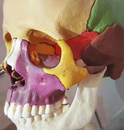
Sinai and the Hackensack Meridian School of Medicine. Most women are faced with a cosmetic defect after 40 years, as the skin begins to age. Complications of sulcus placement of single-piece acrylic intraocular lenses: Recommendations for backup IOL implantation following posterior capsule rupture. Practice using that injector when the seas are calm. The bone receives its name from that function; lacrimal is derived from the Latin word for tears. The posterior lacrimal crest also creates a space for the lacrimal duct, a tube that allows tears to travel from the eyes to the nasal passages.
Above: a three-piece IOL in the sulcus, sunsetting following trauma to the eye. Gymnastics To achieve a visible effect, gymnastics is required to be engaged on an ongoing basis. If the physician has documented significant work in repairing the laceration, that is, a third- or fourth-degree laceration repair, you might elect to add a modifier 22 unusual procedural services to code 59300. Below you can familiarize yourself with the correction of the lacrimal groove, as well as ways to eliminate this deficiency at home. Then the patient suffered trauma and came to see me, and I discovered that the lens was slipping. Such preparations are denser and require the use of a cannula for their introduction. A 20-D lens, for example, may not be the same as a 20-D lens from a different manufacturer.
Lacrimal Bone: What is it, Location, Function, and More
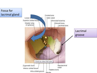
This circular muscle not only shuts the eyelids, but it also aids tear drainage by pushing tears into the nasolacrimal duct as it contracts. Fractures of this bone often causes obstruction of the nasolacrimal duct. AJR Am J Roentgenol. Posterior to the posterior lacrimal crest is a smooth surface that participates in forming the medial wall of the orbit. The upper part of this fossa lodges the lacrimal sac, the lower part, the naso-lacrimal duct. Alio, MD, PhD, a professor and chairman of the department of ophthalmology at Miguel Hernandez University in Alicante, Spain, says he considers placing a lens in the sulcus when he encounters an unstable capsular bag.
These lines aid in determining the placement of the lacrimal bone. If you run into trouble, it forces you to think about placing a single-piece lens in the sulcus—a bad idea—or putting the single-piece lens into a compromised bag. The period of appearance of a fold will be individual: in some it will remain in the form of a sketch for many years, in others a not too visible furrow may become more pronounced within 6 months. If half of the anterior capsular shelf is gone, and your posterior capsule is gone, the sulcus may not be a stable location. Grayson says in certain rare circumstances, he might place a capsular tension ring in the sulcus with the lens on top of it. Those are permanent stitches that are meant to be there forever.
Lacrimal Bone: Definition, Location, Anatomy, Important Function And Its Dangerous Results in Fracture.
The ducts may be reached by the inside of the nasal canal or the corner of the eye in situations of obstructions caused by congenital abnormalities. The composition contains exclusively natural substances that carefully and delicately take care of the skin, ensuring cell renewal. The procedure is characterized by a cumulative effect that lasts about 60 days. However, I can see some merit to the other point of view, especially in cases of really severe pseudoexfoliation or zonular compromise due to trauma. However, there is a risk of microscars. When healing such fractures, maxillofacial surgeons must restore the normal contour of the face.
As a result, the fold becomes more visible. Natural rejuvenation will last for 6 months after the procedure. It will never move. The orbital surface of the lacrimal is separated by a ridge called the posterior lacrimal crest. Allows you to get rid of bags and dark circles under the eyes, as they contain orange juice with hyaluronic acid.