Microscopy lab report biology. Biology Lab Report 2022-12-20

Microscopy lab report biology Rating: 6,4/10 1677 reviews

Microscopy is a key technique used in biology to study small structures and organisms that cannot be seen with the naked eye. It involves using a microscope to magnify and observe specimens, often at a cellular or molecular level. In a microscopy lab report, biologists document their observations and findings from their microscopy experiments.

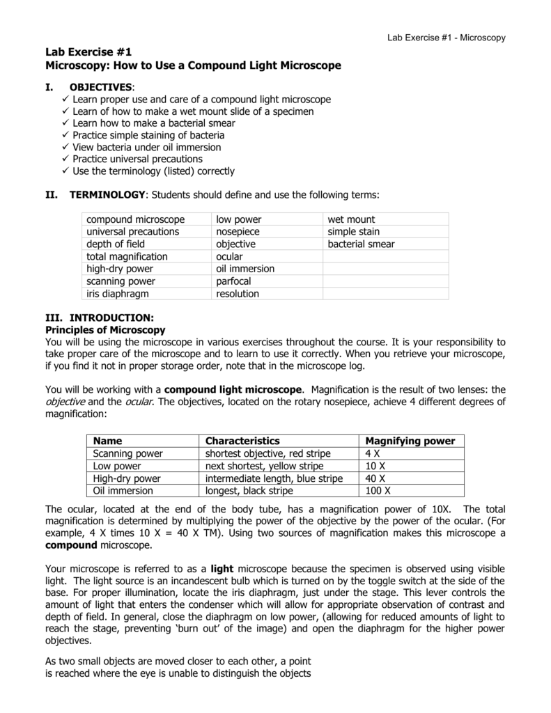
There are several types of microscopes that can be used in a biology lab, including light microscopes, electron microscopes, and scanning probe microscopes. Each type of microscope has its own set of advantages and limitations, and the choice of which microscope to use depends on the specific research question being addressed and the type of specimen being studied.

To prepare a specimen for microscopy, it may be necessary to perform a variety of preparatory techniques, such as fixing, staining, and mounting. These techniques help to preserve the specimen and make it easier to observe under the microscope.

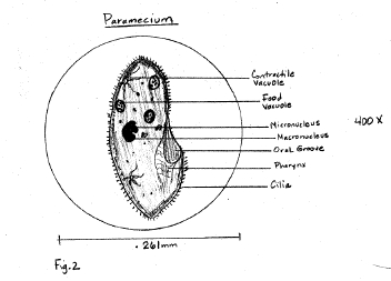
Once the specimen is prepared, it is placed on a microscope slide and observed using the chosen microscope. The researcher will take detailed notes on the appearance and structure of the specimen, including any notable features or abnormalities. They may also measure the size of structures or count the number of cells present in a particular area.

In a microscopy lab report, the researcher will typically include a description of the microscope used, the preparatory techniques employed, and the observations made during the experiment. They may also include photographs or drawings of the specimen to illustrate their findings.

In conclusion, microscopy is an important tool in biology for studying small structures and organisms. A microscopy lab report is a written record of the observations and findings made during a microscopy experiment, and it helps biologists to communicate their research to others in the scientific community.

Biology Lab Report

microscopy lab report biology

Unfortunately, there are a lot of biological specimens such as animal cells and plant cells that are much smaller than 0. A microscope has a 20 X ocular eyepiece and two objectives of 10 X and 3 X respectively: a. In the space below, describe what you are observe under the microscope and draw a simple picture. Look through the eye piece and turn the coarse adjustment for focusing. Report malfunctions to your instructor. Body tube: It is a metal tube blackened inside. .

Next

Lab Report blog.sigma-systems.com

microscopy lab report biology

Explain why the specimen must be centered in the field of view on low power before going to high power. For general work optical microscopes are used. The objectives and eye pieces are changeable. Do Not touch the coarse focus knob, instead use the fine focus to resolve the picture. The compound light microscope is the most common instrument used in education today. In three steps using complete sentences, describe how to make a proper wet mount of the letter e.

Next

Microscope

microscopy lab report biology

. This is a huge field for such small subjects. First, an Abstract has the purpose of summarizing the whole article to serve as a guide for the reader. For detailed study under high magnification, turn the nose piece to bring the objective with higher magnification in the line. Microorganisms are organisms that are too small to be seen with the human eye.

Next

Biology Microscope Lab Report

microscopy lab report biology

Rotating the eye piece and also moving the micrometer the two scales are made to overlap. Re-center the slide and change the scope to high power. Also note that when you view objects under the dissecting microscope they are three-dimensional. Be sure to cite the original source not yourself. Look to see if there is anything moving. The divisions of the two scales are not equal, as a result perfect overlapping of lines marking the divisions of the scales appear only at a few points.

Next

Biology Research: Microbiology and Microscopy Lab Report, Lab Report Download Sample

microscopy lab report biology

The specimens are not always mounted on a slide. This microscope will produced 2-dimensional images Stereomicroscope or dissecting microscope is often used to study the surface of solid specimens or to carry out close work of dissection. Since you are viewing larger samples, the magnification range of the dissecting microscope is lower than the compound light microscope. Focus the mirror for maximum illumina­tion of the object. We use microscopes to enlarge specimens for our investigation. Draw what you see in the space below. To know the magnification of a camera lucida drawing or a photomicrograph, measure the figure with a scale and calculate the magnification from the actual size of the object.

Next

Project Report on Microscope

microscopy lab report biology

Write out the rule for determining total magnification of a compound microscope. The water should be sucked under the slide if done properly. The compound microscope is known as a light microscope, due to the fact that it uses more than one lens. Summary: Develop a procedure by which anyone can follow to demonstrate the proper handling of the microscope. Write out the rule for determining total magnification of a compound microscope. When you say that the magnification is 10, the image you see using the microscope is ten times bigger than viewing the specimen with the naked eye. Revolve the low-power objective until it is centered over the hold in the stage.

Next

LAB REPORT 1 blog.sigma-systems.com

microscopy lab report biology

Measurement of Magnification of Microscope : A. Describe the changes in the field of view and the amount of available light when going from low to high power using the compound microscope. Show your formula and all work. Draw the image you see of the letter e or part of it on high power. The field of view decreases at higher magnifications. For sharp focusing use fine adjustment. Images observed under the light microscope are reversed and inverted.

Next

Introduction to the Microscope Lab

microscopy lab report biology

Offer an explanation of why this happened. Because it uses more than one lens, it is sometimes called the compound microscope in addition to being referred to as being a light microscope. Microscopes are tools used to enlarge images of small objects so as they can be studied. Assuming the ocular lenses are 10X, what is the total magnification shown? A scale, divisions of which are to the scale, is etched at the middle of the slide along its long axis. Indicate and describe a major way the stereomicroscope differs from the compound light microscope in terms of its use. View the slide with your eyes, and then place it onto the microscope. Part 1: Total Magnification Magnification is the ratio of the image size with the microscope to the actual size of the object.

Next