Most superficial tunic. Blood Vessel Layers: Tunica Intima, Tunica Media & Tunica Adventitia 2022-12-10
Most superficial tunic
Rating:
8,8/10
1307
reviews
The most superficial tunic, also known as the epidermis, is the outermost layer of the skin. It is a thin, protective layer that helps to shield the body from environmental damage and infection. The epidermis is made up of multiple layers of skin cells, and it is constantly shedding and replenishing itself.
One of the main functions of the epidermis is to act as a barrier against external factors such as bacteria, toxins, and UV radiation. It is composed of a type of protein called keratin, which helps to keep the skin strong and resistant to damage. The epidermis also contains melanocytes, which produce the pigment melanin that gives skin its color.
The epidermis is a dynamic layer of the skin, constantly undergoing changes in response to various internal and external stimuli. It is regulated by hormones, such as estrogen and testosterone, and can be affected by a variety of factors including stress, diet, and genetics.
Despite its importance in protecting the body, the epidermis is often thought of as the most superficial layer of the skin because it is the outermost layer that we see. However, it is worth noting that the epidermis plays a vital role in maintaining overall skin health and function.
In conclusion, the epidermis, or most superficial tunic, is a crucial layer of the skin that helps to protect the body from external damage and infection. It is constantly shedding and replenishing itself in response to various stimuli, and it plays a vital role in maintaining overall skin health.
What is the longest superficial vein in the body?

Transition to the sclera, with more distinct fibrillation, and surmounted by a thicker epithelium. Corneal corpuscles appearing fusiform in section. What are the three layers of the neural tunic of the eye wall quizlet? The cone granules, fewer in number than the rod granules, are placed close to the membrana limitans externa, through which they are continuous with the cones of the layer of rods and cones. The muscular fibers are involuntary, and consist of circular and radiating fibers. What is the sclera and its function? Again, the iris may be the seat of a malformation, termed coloboma, which consists in a deficiency or cleft, clearly due in a great number of cases to an arrest in development.
Next
Exercises 27, 29

The Ciliary Body corpus ciliare. Their posterior surfaces are covered by a bilaminar layer of black pigment cells, which is continued forward from the retina, and is named the pars ciliaris retinæ. The thinnest area of the human sclera is approximately 0. The ciliary processes processus ciliares are formed by the inward folding of the various layers of the choroid, i. Immediately after the second heart sound, both the 6 atria and 7 ventricles are filling with blood. Internal to this lamina is the choroid proper, consisting of two layers: an outer, composed of small arteries and veins, with pigment cells interspersed between them; and an inner, consisting of a capillary plexus. At the fovea centralis the retina is exceedingly thin, and the dark color of the choroid is distinctly seen through it.
Next
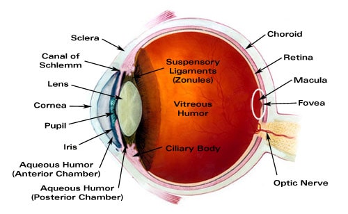
What are the 3 layers of the eye?
These lamellæ are made up of bundles of modified connective tissue, the fibers of which are directly continuous with those of the sclera. Pingueculae do not go away on their own and do not require treatment in most cases. When viewed from the outer surface these cells are smooth and hexagonal in shape; when seen in section each cell consists of an outer non-pigmented part containing a large oval nucleus and an inner pigmented portion which extends as a series of straight thread-like processes between the rods, this being especially the case when the eye is exposed to light. To help stand up to the high demand, the walls of the blood vessels are constructed of three layers known as tunics. Circulus iridis major cut across.
Next
A&P 2 Blood Vessels Flashcards

A pinguecula is yellowish in color and typically has a triangular shape. Choroid: A layer of connective tissue between the retina and sclera. The choroid invests the posterior five-sixths of the bulb, and extends as far forward as the ora serrata of the retina. Their posterior surfaces are connected with the suspensory ligament of the lens. Course of vasa ciliar. The capillary net-work close to the pupillary margin of the iris.
Next
The Tunics of the Eye
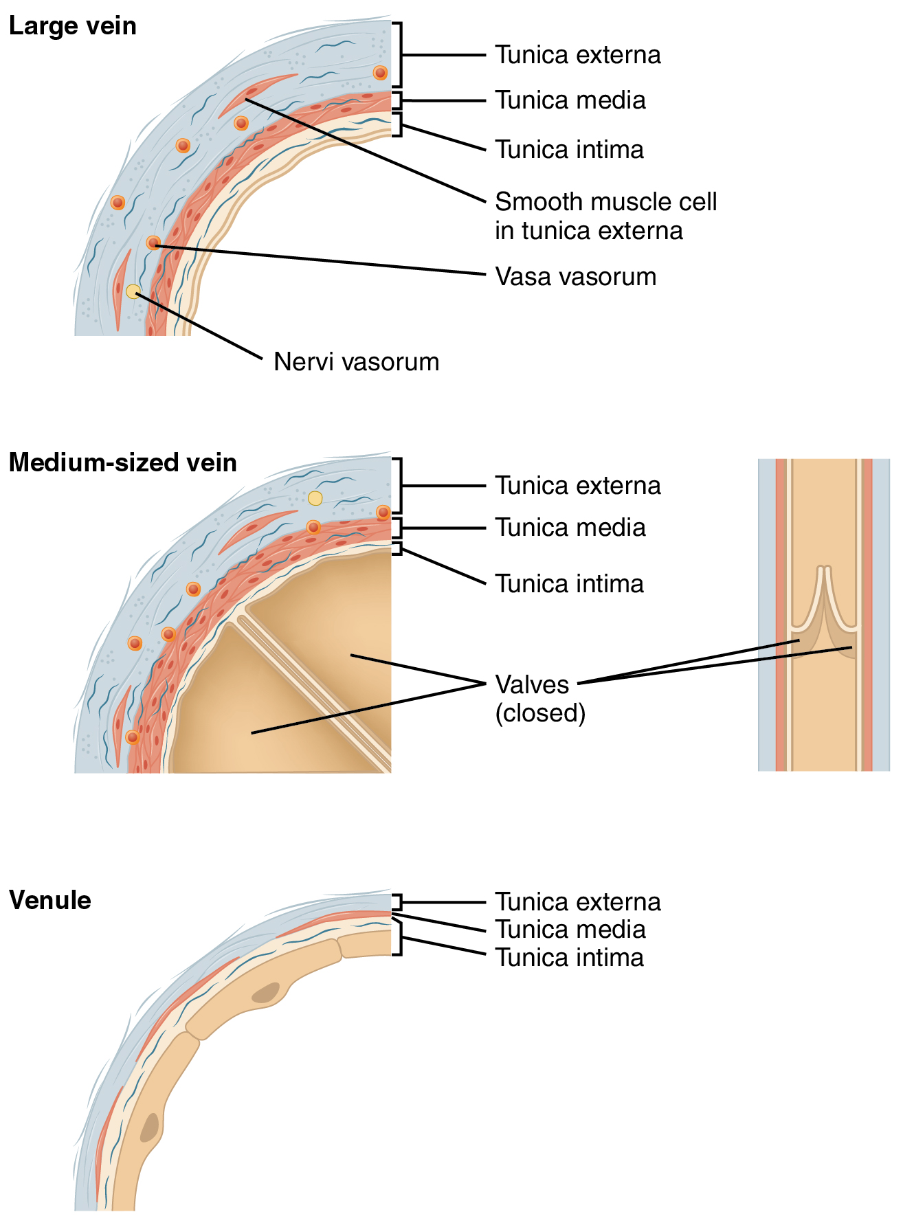
Lastly, there are three or four layers of squamous cells, with flattened nuclei. They are divisible into rod bipolars and cone bipolars. It also contains strong collagen fibers that help anchor the blood vessel to surrounding tissues, and this gives the vessel some stability. First, light passes through the cornea the clear front layer of the eye. The cones are conical or flask-shaped, their broad ends resting upon the membrana limitans externa, the narrow-pointed extremity being turned to the choroid. Where the optic nerve passes through the sclera, the latter forms a thin cribriform lamina, the lamina cribrosa scleræ; the minute orifices in this lamina serve for the transmission of the nervous filaments, and the fibrous septa dividing them from one another are continuous with the membranous processes which separate the bundles of nerve fibers. The choriod is filled with blood vessels that bring oxygen and nutrients to the eye.
Next
Blood Vessel Layers: Tunica Intima, Tunica Media & Tunica Adventitia
Each begins on the inner surface of the retina by an expanded, often forked base, which sometimes contains a spheroidal body staining deeply with hematoxylin, the edges of the bases of adjoining fibers being united to form the membrana limitans interna. What are the three layers of the eye? Suspensory ligament of lens. The cells are somewhat flask-shaped; the rounded internal surface of each resting on the stratum opticum, and sending off an axon which is prolonged into it. The macula receives two small branches superior and inferior macular arteries from the temporal branches and small twigs directly from the central artery; these do not, however, reach as far as the fovea centralis, which has no bloodvessels. Identify each; and on the lines to the sides, note the structural details that enabled you to make these identifications? It may be regarded as a condensed part of the substantia propria. The outer layer lamina vasculosa consists, in part, of the larger branches of the short ciliary arteries which run forward between the veins, before they bend inward to end in the capillaries, but is formed principally of veins, named, from their arrangement, the venæ vorticosæ. This is termed the subepithelial plexus, and from it fibrils are given off which ramify between the epithelial cells, forming an intraepithelial plexus.
Next

They are well-developed in hypermetropic, but are rudimentary or absent in myopic eyes. After reaching the iris they form a plexus around its attached margin; from this are derived non-medullated fibers which end in the Sphincter and Dilatator pupillæ their exact mode of termination has not been ascertained. Where is sclera located in the eye? The cornea and the lens help to focus the light rays onto the back of the eye retina. Behind it is pierced by the optic nerve, and is continuous through the fibrous sheath of this nerve with the dura mater. Arteriæ, and I 1. What holds the eyeball in place? In these cases the cleft is found at the lower aspect, extending directly downward from the pupil, and the gap frequently extends through the choroid to the porus opticus.
Next
Its outer surface is in contact with the choroid; its inner with the hyaloid membrane of the vitreous body. Veins Which depends on elasticity to propel blood along; arteries or veins? The meridional fibers, much the more numerous, arise from the posterior margin of the scleral spur page 1007 ; they run backward, and are attached to the ciliary processes and orbiculus ciliaris. The chemical and optical characters of the two portions are identical with those of the rods. Anastomosis with branches of short posterior ciliary arteries. This double layer is known as the pars ciliaris retinæ, and can be traced forward from the ciliary processes on to the back of the iris, where it is termed the pars iridica retinæ or uvea. The iris may be absent, either in part or altogether as a congenital condition, and in some instances the pupillary membrane may remain persistent, though it is rarely complete.
Next

The front part of the choroid has two structures: The ciliary body, a muscular area attached to the lens. You can easily recall this by remembering that 'media' and 'middle' and 'muscular' all start with the same letter, which is 'm. The tunica adventitia layer is made up of connective tissue and withstands outside forces Lesson Summary Let's review. Veins are blood vessels that carry blood towards the heart. Oblique fibers in the anterior layer of the substantia propria.
Next