Pterygoid bone. Pterygoid Definition & Meaning 2022-12-27
Pterygoid bone Rating:
7,2/10
692
reviews
The pterygoid bones, also known as the pterygoid processes, are a pair of bones located in the human skull. These bones are located just below the eye sockets and are a part of the sphenoid bone, which forms the central base of the skull. The pterygoid bones are elongated and triangular in shape, and they are connected to the sphenoid bone by a thin, bony structure known as the pterygoid lamina.
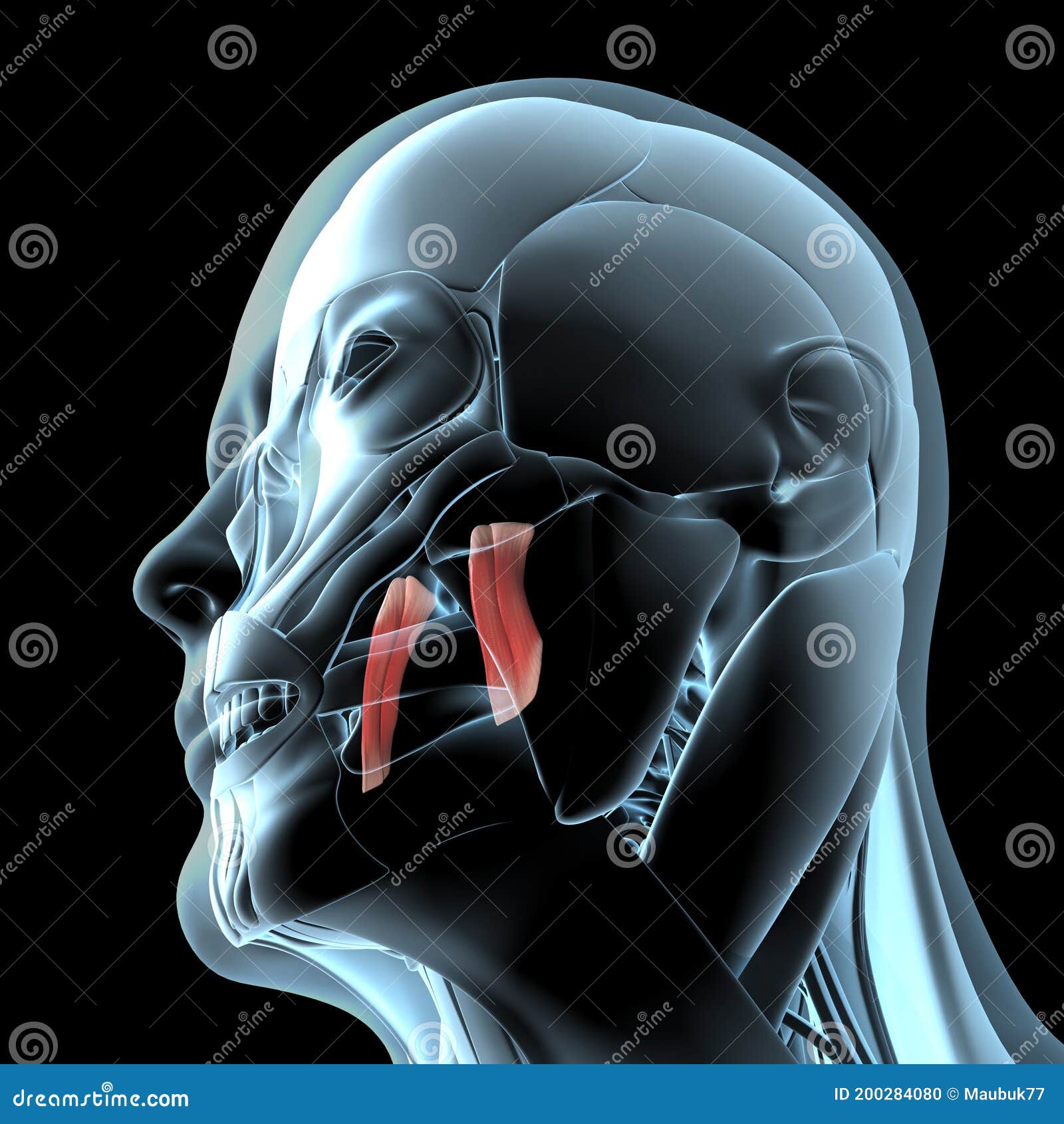
The primary function of the pterygoid bones is to support the muscles of the jaw and to assist in the movement of the mandible, or lower jaw. The pterygoid bones are connected to several muscles that help to move the jaw forward, backward, and side to side, allowing us to chew and speak. In addition to their role in jaw movement, the pterygoid bones also provide support for the nasal cavity and the nasal passages.
The pterygoid bones are made up of three main parts: the pterygoid process, the pterygoid fossa, and the pterygoid hamulus. The pterygoid process is a bony protrusion that extends from the sphenoid bone and is responsible for attaching several jaw muscles. The pterygoid fossa is a depression located on the inner surface of the pterygoid bone and is used for the attachment of various muscles and ligaments. The pterygoid hamulus is a small, hook-like structure that is located on the outer surface of the pterygoid bone and is used for the attachment of the tensor veli palatini muscle, which helps to elevate the palate during swallowing.
In addition to their role in supporting the muscles of the jaw and the nasal passages, the pterygoid bones are also involved in the process of breathing. The pterygoid muscles, which are connected to the pterygoid bones, play a role in regulating the airflow through the nasal passages. Dysfunction of the pterygoid muscles can lead to breathing difficulties and may contribute to conditions such as sleep apnea.
Overall, the pterygoid bones are an important part of the human skull, playing a vital role in the movement of the jaw and the regulation of breathing. They provide support for several muscles and assist in the movement of the mandible, allowing us to chew and speak. They also contribute to the overall structure and function of the nasal cavity and nasal passages.
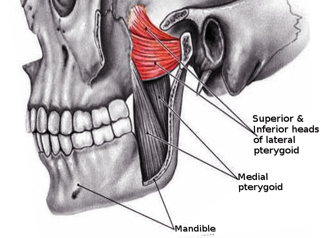
Medial and lateral pterygoid muscle: Anatomy and function
The slow worm Anguis fragilis is a legless lizard that can grow to a length of 50cm and superficially resembles a snake. The primary function of the lateral pterygoid muscle is to pull the head of the condyle out of the mandibular fossa along the articular eminence to protrude the mandible. It is usually easily palpable. While patients will always have the option to be fitted with dentures or a dental partial, dental implants offer the best long-term solution for missing teeth. It connects to the middle and inner ear cavities of the temporal bone. In its rostral part the vomer is shaped, in vertical section, like a Y or perhaps a T. The occipital bone forms the back of the skull.
New York: Thieme Medical Publishers, Inc. Emi Knafo, in Mader's Reptile and Amphibian Medicine and Surgery Third Edition , 2019 Crocodilians. Schedule a free consultation with Dr, Mike at Cleveland Implant Institute to learn more about this exciting new procedure. Musculus pterygoideus medialis Musculus pterygoideus internus Medial pterygoid is a thick quadrilateral muscle that connects the Medial pterygoid muscle consists of two heads; superficial and deep. Available under CC-BY-SA license. Most sounds are heard by our eardrums.
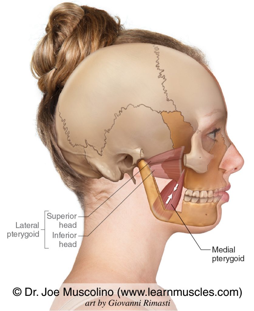
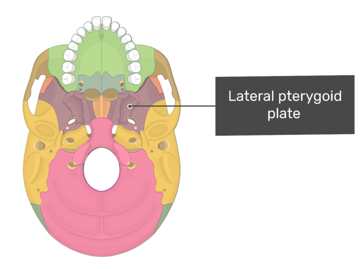
Our culture values male pleasure more than female pleasure. Key facts of the medial pterygoid muscle Origin Superficial part: Tuberosity of maxilla, Pyramidal process of palatine bone; Deep part: Medial surface of lateral pterygoid plate of sphenoid bone Insertion Medial surface of ramus and angle of mandible Action Bilateral contraction - Elevates and protrudes mandible Unilateral contraction - Medial movement rotation of mandible Innervation Medial pterygoid nerve of mandibular nerve CN V3 Blood supply Pterygoid branches maxillary artery, buccal artery, facial artery This article will discuss the Synonyms: Deep part of medial pterygoid muscle, Pars profunda musculi pterygoidei medialis Medial pterygoid muscle consists of two heads; superficial and deep, that are separated by the inferior head of lateral pterygoid muscle at their origin. They are cold-blooded reptiles. Snakes have between 200-400 vertebrae with as many ribs attached. Kenhub does not provide medical advice. In case you were wondering cause they are soooo flexible , snakes actually do have bones. These include the bones of the skull, spine vertebrae , ribs, arms and legs.
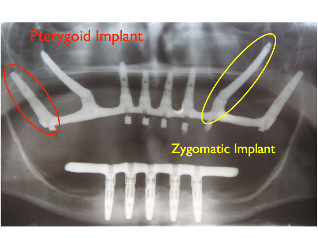
This space is largely filled by the ethmoturbinates, complex bony processes that arise from the ethmoid. I only use these implants when I have to. I often use them in combination with A pterygoid implant cannot stand alone. The latter, forming the bottom of the burr slot, is identified when the red cancellous layer is replaced by a white inner cortical table. The information we provide is grounded on academic literature and peer-reviewed research.
Why is it called pterygoid? When snakes are kept starved for days and offered milk, they do drink to keep them hydrated. After the functional tooth is shed, the replacing tooth moves into position while still only about one-third of its final size. How do you pronounce pterygoid? All content published on Kenhub is reviewed by medical and anatomy experts. Conversely, pterygoid implants do not necessitate sinus augmentation, therefore patients can enjoy their new smile in less time. Is pterygoid a skull bone? It is located in the most inferior portion of the cranial fossa as a part of the occipital bone.
The first maxillary tooth is larger than the premaxillary teeth and the size increases to a maximum in the fifth or sixth position, decreasing again posteriorly. At birth, there are 9 premaxillary teeth, including a median tooth, 10 on each maxilla and 11 on each dentary. They almost reach into the wisdom teeth areas. The ethmoid bone, located at the roof of the nose between the eye sockets, separates the nasal cavity from the brain. This also supplies the tensor tympani muscle and the tensor veli palatini muscle. Towards the back the long stem of the T disappears and is replaced by cartilage. Consequently, a traumatic blow to the pterion may rupture the middle meningeal artery causing an epidural haematoma.
Start with the muscles of mastication by exploring our videos, quizzes, labelled diagrams and articles. Although having different origins, both heads insert on the inner surface of mandible, creating an axis for a strong pull of this bone. By the second year of life, there are still 9 teeth on the premaxillae, but only 9 on each maxilla and 10 on each dentary. WHERE DOES A PTERYGOID IMPLANT GO? The pterygoid implant goes where your wisdom teeth on the upper jaw used to be. Textbook of Anatomy Regional and Clinical Head, Neck, and Brain; Volume III. Unilateral contraction of medial pterygoid causes rotation of mandible, while bilateral contraction elevates and protrudes it.
The femoral head is prominent and deviates medially from the shaft. What is an Anatolian bump? What is an Inca bone? What is the pterygoid canal? Also the implants are placed sometimes partially in and partially out of the sinuses similar to some of the zygomatic implants that I place. The ethmoid bone is completely hidden by the maxilla and nasal bones and part of the frontal bones , indeed the ethmoid and maxilla form a double wall, which we shall return to when we describe the maxillary sinus of the rat. The foramen magnum is the largest foramen of the skull. The fibers attach via a strong tendinous lamina that extends from the Take this quiz to test yourself on the muscles of mastication. In the rat the vomer, in the midline, and a pair of shelf-like plates from the maxilla to the front, and from the ethmoid to the back, form the floor of the posterior part of the nasal cavity.