Right common iliac artery. Iliac Artery 2022-12-26

Right common iliac artery Rating: 7,7/10 809 reviews

The right common iliac artery is a major blood vessel in the human body that carries oxygenated blood from the aorta to the lower half of the body. It is one of two common iliac arteries, the other being the left common iliac artery, which carries blood to the left side of the body.

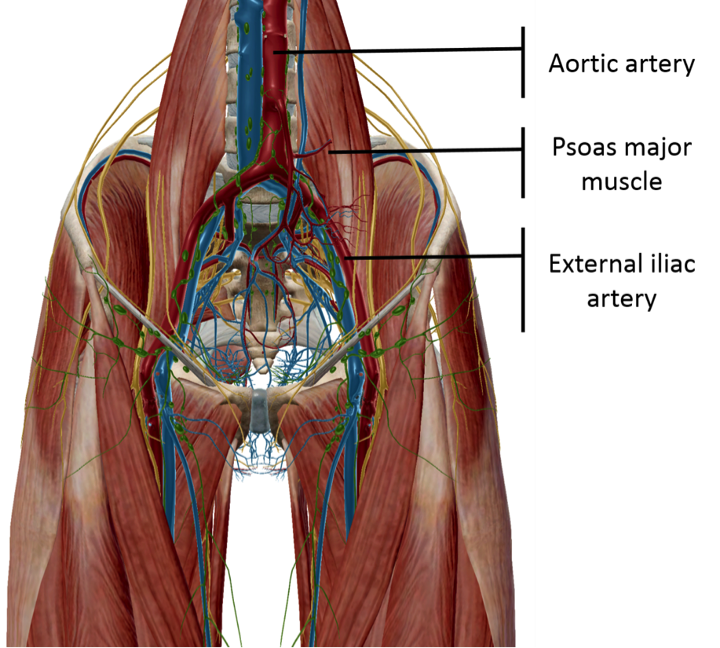
The right common iliac artery begins at the level of the fourth lumbar vertebra, where it branches off from the aorta. From there, it travels downward and to the right, passing through the pelvis and into the right leg. Along its course, the right common iliac artery gives off several smaller branches, including the internal iliac artery and the external iliac artery.

The internal iliac artery supplies blood to the muscles and organs of the pelvis, including the bladder, uterus, and rectum. The external iliac artery, on the other hand, supplies blood to the muscles and skin of the lower extremities, including the thighs, knees, and ankles.

Problems with the right common iliac artery can have serious consequences for a person's health. One such problem is an arterial blockage, also known as arterial occlusion, which can lead to reduced blood flow to the lower half of the body. This can cause symptoms such as leg pain, numbness, and weakness, and if left untreated, it can lead to more serious complications such as tissue death and amputation.

Treatment for problems with the right common iliac artery typically involves the use of medications to widen the artery and improve blood flow, as well as lifestyle changes such as quitting smoking and adopting a healthy diet. In more severe cases, surgical procedures may be necessary to repair or bypass the damaged artery.

In conclusion, the right common iliac artery is a vital blood vessel that plays a crucial role in supplying oxygenated blood to the lower half of the body. Problems with this artery can have serious consequences for a person's health, but with proper treatment, it is possible to manage and prevent these problems from occurring.

Iliac artery aneurysm

right common iliac artery

Anatomy, Abdomen and Pelvis, External Iliac Arteries. Rupture is associated with a 58% mortality 10. Aneurysms in the brain may expand without breaking open rupturing. The ovaries receive their blood supply primarily from the aorta. Solitary aneurysms of the iliac arterial system: an estimate of their frequency of occurrence.

Next

Common Iliac Artery: Anatomy, Function, and Significance

right common iliac artery

The ascending aorta performs the function of supplying blood to the head, neck, and the arms, whereas the thoracic aorta supplies blood to the organs located in the chest cavity. About 13,000 Americans die each year from aortic aneurysms. Popliteal artery Descending genicular artery Superior medial genicular artery Inferior medial genicular artery Top Anterior view of the arteries of the thigh is shown. Aortic angiography shows the inside of your aorta. The graft is then expanded inside the aorta and fastened in place to form a stable channel for blood flow. The inferior gluteal vessels pass through the lower part of the greater sciatic foramen. It is important to maintain the health of this artery in order to prevent serious complications such as reduced blood flow and the development of an aneurysm.

Next

Common Iliac Artery Anatomy, Function & Diagram

right common iliac artery

These arteries carry blood to the pelvis and lower limbs. These two arteries travel down and to each side of the body for about five centimeters towards the edges of the pelvis. It supplies blood to the psoas and quadratus lumborum. The obturator nerve mimics the course and divisions of the obturator vessels. Both common iliac arteries branch from the base of the aorta. A vascular surgeon does surgery on the aorta and other blood vessels, except those of the heart and brain.

Next

Iliac Artery

right common iliac artery

It crosses behind the right common iliac artery before joining the right vein to form the inferior vena cava Fig. On any matter relating to your health or well-being, please check with an appropriate health professional. The uterine artery branches into arcuate arteries in the uterine wall. Conditions and Disorders What conditions and disorders affect the iliac arteries? The carotid arteries are the two main arteries on each side of your neck. They form the inferior vena cava. Therefore, they should be routinely identified and protected during peripheral compartment arthroscopy and during open joint-preserving hip surgery.

Next

Guidance for Surveillance of Isolated Common Iliac Artery and Small Abdominal Aortic Aneurysms

right common iliac artery

Palpation of the sciatic notch and the posterior column can help to prevent the placement of proud screws and further decrease the risk of injury. Medicines If you have an aortic aneurysm, your doctor may prescribe medicines before surgery or instead of surgery. Anatomy of iliac arteries The aorta divides into the right and left common iliac arteries when it reaches the fourth vertebra of the spine. Feasibility of a branched stent-graft in common iliac artery aneurysms. When they do occur, it is usually as a result of anteriorly placed deep retractors or during cement extrusion in this region. Atlas of Human Anatomy 7th ed. What problems affect the iliac arteries? These vessels supply blood to the muscles and skin in the lower abdominal wall.


Next

External Iliac Artery: Anatomy, Function, Significance

right common iliac artery

What do the internal iliac arteries supply? Last medically reviewed on January 19, 2018. Superficial circumflex iliac artery Lateral circumflex femoral artery Perforating vessels Descending branch lateral circumflex femoral artery Superior lateral genicular artery Inferior lateral genicular artery Internal iliac artery Obturator artery Inferior epigastric a. Smoking can damage and weaken the walls of the aorta. There are also surgical options like minimally invasive endovascular techniques, stent-graft, or open surgery that can be used to repair the aneurysm. These are also paired, which means that they are present on the left, as well as the right. The femoral artery continue into the leg to become the popliteal artery behind the knee, and the anterior and posterior tibial arteries in the legs. Signs and symptoms also depend on whether the aneurysm has ruptured burst or is affecting other parts of the body.

Next

Anatomy and Function of the Common Iliac Artery With Labeled Diagrams

right common iliac artery

Isolated iliac artery aneurysms are rare clinical conditions 3. The common iliac veins formed by the union of external and internal iliac veins lie medial to the corresponding arteries at a deeper plane. If iatrogenic injury does occur, the external iliac artery and vein can be accessed most easily through the ilioinguinal approach. Injuries to the profundus femoris and its branches have been described during arthroplasty approaches to the hip, but they are fairly unusual. These tests can show the size of an aortic aneurysm, if one is found. Mohan Garikiparithi got his degree in medicine from Osmania University University of Health Sciences. Iliac artery aneurysm An aneurysm is an abnormal widening or ballooning of a part of an artery due to weakness in the wall of the blood vessel 1.

Next

Common iliac vein: Anatomy and drainage

right common iliac artery

In case of an anatomical variation where the common iliac arteries are absent, the external and internal iliac arteries will arise together from the distal end of the aorta. The lateral sacral artery gives off a superior and inferior branch. Guidance for Surveillance of Isolated Common Iliac Artery and Small Abdominal Aortic Aneurysms The common iliac arteries originate near the fourth lumbar vertebra in the lower back, where the abdominal aorta divides bifurcation. The largest artery in the body the aorta divides into the common iliac arteries. Trauma, such as a car accident, also can damage the walls of the aorta and lead to thoracic aortic aneurysms. Only the proximal portion of the umbilical arteries remains patent after birth, while the distal portion obliterates forming the medial umbilical ligaments. Anatomy of the femoropopliteal artery and its branches.

Next

Iliac Artery: What Is It, Location, Anatomy and Function

right common iliac artery

Fibromuscular dysplasia Narrowing and enlargement of the medium-sized arteries can be associated with Cystic medial necrosis When there is damage to connective tissues of large arteries, this is known as cystic medial necrosis. Erroneous posterior screw placement can cause the disruption of these structures. It often takes a month to recover from open abdominal or open chest surgery and return to full activity. Diagnosis of iliac artery aneurysm There is no commonly accepted way to detect iliac artery aneurysms. Aneurysms can develop and grow large before causing any signs or symptoms.


Next