Scanning electron microscope how it works. What is Scanning Electron Microscopy? (How it Works, Applications, and Limitations) 2023-01-07
Scanning electron microscope how it works Rating:
4,7/10
798
reviews
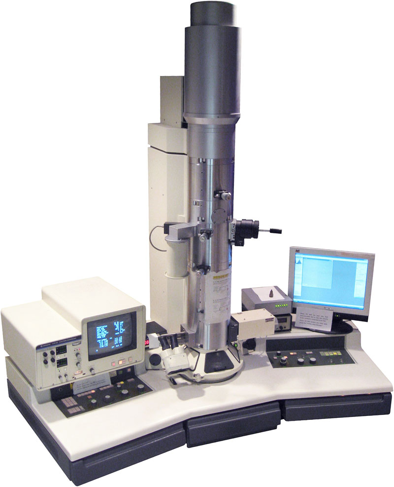
A scanning electron microscope (SEM) is a type of microscope that uses a beam of electrons to create an image of a sample. SEMs are commonly used in scientific research and industrial applications to analyze the surface structure and composition of materials at a high level of detail.
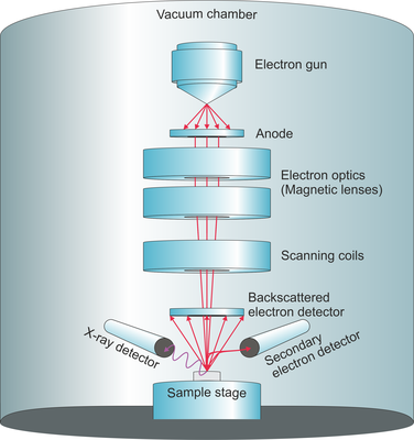
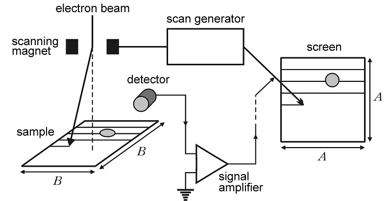
To use a SEM, the sample is placed on a stage and a beam of electrons is emitted from a filament and focused onto the sample using a series of lenses. As the electrons interact with the sample, they can knock other electrons out of the sample's surface, a process called secondary electron emission. These secondary electrons are collected by a detector and used to create an image of the sample.
In addition to secondary electrons, the interaction of the electron beam with the sample can also produce other types of signals, such as backscattered electrons and X-rays. These signals can provide additional information about the sample, such as its elemental composition and crystal structure.
One of the major advantages of SEMs is their ability to produce high resolution images of samples, with magnifications up to several hundred thousand times the original size. This high resolution allows scientists and researchers to analyze the surface of materials at a level of detail that is not possible with other types of microscopes.
SEMs are also capable of operating in a variety of modes, such as high-angle annular dark field (HAADF) mode and energy dispersive X-ray spectroscopy (EDS) mode, which allow for the analysis of different aspects of the sample.
In summary, the scanning electron microscope is a powerful tool that allows scientists and researchers to analyze the surface structure and composition of materials at a high level of detail. Its ability to produce high resolution images and operate in a variety of modes makes it a valuable tool for a wide range of applications.
Scanning electron microscopy (SEM), what is it for?
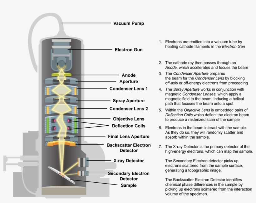
WhatCreates the Electrons Used in SEM? Minimal preparation includes acquisition of a sample that will fit into the SEM chamber and some accommodation to prevent charge build-up on electrically insulating samples. Ever wonder how we can see things so small, the human eye doesn't even know it exists? That's where a scanning electron microscope SEM came in. Press and hold the VENT button until it flashes and you will hear a click. Zeitschrift für Technische Physik. This is extremely important because the chamber of the SEM needs to stay very clean! Journal of Electron Microscopy Technique. This image also shows that as the beam penetrates the sample, it widens.
What is Scanning Electron Microscopy? (How it Works, Applications, and Limitations)
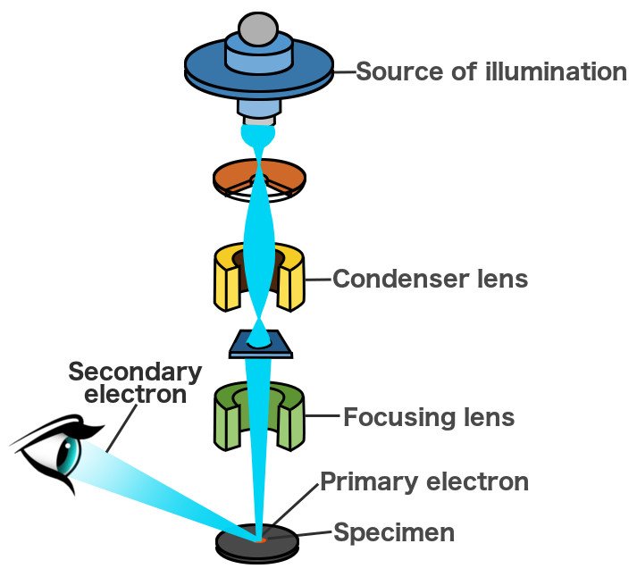
The SEM's job is to use an electron beam to trace over the object, creating an exact replica of the original object on a As the electron beam traces over the object, it interacts with the surface of the object, dislodging secondary electrons from the surface of the specimen in unique patterns. CRC Press electronic resource Related Links For more information about Scanning Electron Microscopy SEM follow the links below. Electrons are minute-charged particles that occupy the outer regions of atoms. You can detect what elements are present in the examined sample, and even create maps to figure out the composition of precipitates. Instead of increasing temperature, a very high voltage is applied to the cathode. Essentially, an electron beam is generated, focused, and then used to scan across a sample in a raster pattern.
A Brief Introduction to SEM (Scanning Electron Microscopy)
The objective lens and aperture provide the final focus between the electron beam and the sample. Today I'll be writing about how scanning electron microscopes allow us to see far beyond what traditional microscopes can provide. SE images contain more detailed surface information. The choice of material for conductive coatings depends on the data to be acquired: carbon is most desirable if elemental analysis is a priority, while metal coatings are most effective for high resolution electron imaging applications. BSE images provide valuable crystallographic, topographic and magnetic field information.
Tell us about it on our networks, write to us at Related posts: Related projects:. Now press and hold the VENT button until it blinks to return the sample chamber to atmospheric pressure. They are then accelerated and attracted by the positively-charged anode. The electron beam passes through a number of electromagnetic lenses to demagnify and focus the beam, allowing a higher resolution for more detailed imaging. In this article, we'll learn how SEMs are able to produce such detailed and striking images. How Scanning Electron Microscopes Works In scanning electron microscopes SEMs , the electron beam scans the sample in a raster pattern. Hard, dry materials such as wood, bone, feathers, dried insects, or shells including egg shells Fixation is usually performed by incubation in a solution of a The dry specimen is usually mounted on a specimen stub using an adhesive such as epoxy resin or electrically conductive double-sided adhesive tape, and If the SEM is equipped with a cold stage for cryo microscopy, Freeze-fracturing, freeze-etch or freeze-and-break is a preparation method particularly useful for examining lipid membranes and their incorporated proteins in "face on" view.
This creates an electric field which propels the electrons into a beam which shoots through the microscope. They simply consist of coils of wires inside metal pole pieces. Types of Electron Microscopes Transmission Electron Microscope TEM A transmission electron microscope TEM is also known as the original form of the electron microscope. They are then accelerated and attracted to the positively charged anode. Electrons try to move out of the specimen and jump across the gap into the probe. Besides protecting the electron source from being contaminated, vacuum also allows the user to acquire a high-resolution image. The ATRIA materials team explains it to you in this post! The only requirement that the use of this technology implies is that the sample must be conductive, since obtaining the image is the product of the interaction of the electrons emitted by the equipment and the sample.
Does a defect appear continuously, and would you like to know what it is due to? So you will turn it to about 19mm. They need to be conductive, which can be achieved by applying a thin film of metal such as gold. When the high energy electron beam strikes the surface, it has the ability to eject an electron resident in the material from its shell. High-quality 2D SEM images, on the other hand, are widely available. Non-metal samples are trickier to use in SEM. Ga + ions are 130,000 heavier than electrons, so the interaction with the sample is stronger although its penetration is less. The copy isn't made all at once, but rather traced out from one end to the other.
BSEs and SEs contain different types of information. Discover More AboutSEM Please call us on +44 0 161 442 9963, email At SciMed we are keen to educate our viewers with the latest product videos, event updates and even testimonials from our customers! The electrons interact with atoms on the surface of the sample. Once it is in focus, your picture is ready! Whatever the original working distance was, subtract 10 from is and this is how much you will need to move your Z-knob. However, it is also possible to find dedicated STEMs. Samples likely to outgas at low pressures rocks saturated with hydrocarbons, "wet" samples such as coal, organic materials or swelling clays, and samples likely to decrepitate at low pressure are unsuitable for examination in conventional SEM's. As these beams are very focused, they allow better resolution. The main limitation of SEM is the sample preparation.
This part can be tricky. In conclusion, we obtain a high resolution image of the surface topography of our sample. Journal of Vacuum Science and Technology. Otherwise, it's coated in a conductive material like gold or platinum through a process called sputter coating before it's ready for viewing. Samples will always need to be dehydrated, however. Preparing a sample for electron microscopy is a very in-depth procedure on its own, so for this procedure, use a sample that has been prepared for you. .
How is Electron Microscopy Different to Optical Microscopy? It is used to analyse filament bulbs at traffic accident sites. Since these electrons are produced deeper inside the sample, they may exit at the Bragg condition, which provides information on the spacing between atoms. The images obtained have a high resolution. Any JEOL microscope will have extremely similar controls to the microscope described in these instructions. Forensics SEM is a reliable method for analysing gunshot residue, and for analysing paint particles and fibres at crime scenes. Take note of this number. X-ray signals at each position in the scanning area may be compiled to generate a compositional map of the area, which can be useful when identifying phases within a material.
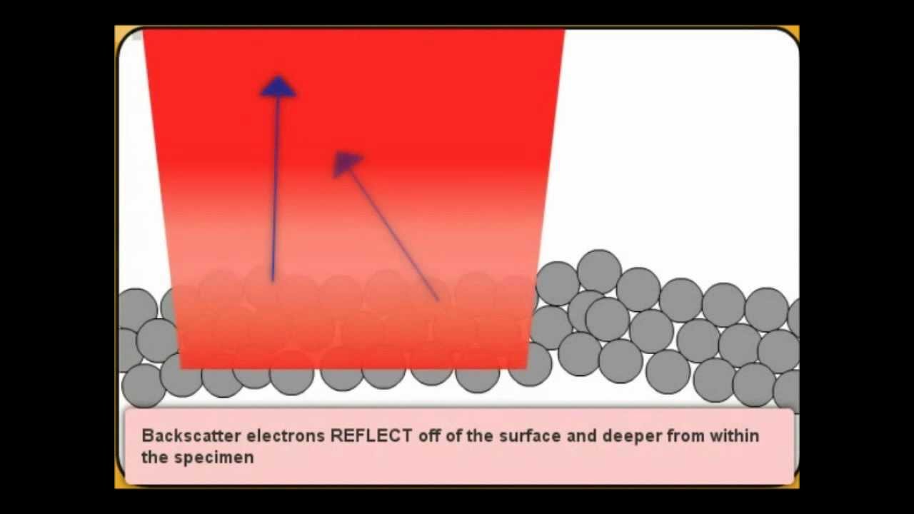
Backscattered and secondary electrons The interaction of electrons within a sample can generate many different types of electrons, photons, or irradiations. By separating the samples from the vacuum chamber using a high-strength film, these SEMs can observe objects never before subject to such high levels of magnification. Although this energy packet is usually a photon, sometimes it may be an electron. New York: Plenum Press. SEM is an extremely versatile tool which is relatively easy to learn and to prepare samples for, compared to something like TEM or atom probe tomography. Thus, the emitted x-rays can be used to identify the different atoms which make up a material. Now your sample is in focus, but you might notice it doesn't look as good as you would expect.