Sensory and motor pathways. 12.7E: Sensory and Motor Tracts 2023-01-01
Sensory and motor pathways Rating:
7,1/10
1695
reviews
The sensory and motor pathways are the means by which information is transmitted between the body and the brain. They allow us to perceive the world around us, to respond to stimuli, and to control our movements.
The sensory pathways begin with sensory receptors, which are specialized cells that detect stimuli such as light, sound, and touch. These receptors send signals through the sensory nerves to the spinal cord and brain, where the information is processed and interpreted.
There are several different types of sensory receptors, each of which is sensitive to a specific type of stimulus. For example, photoreceptors in the retina of the eye detect light, while auditory receptors in the ear detect sound waves. Other types of sensory receptors include those that detect temperature, pressure, and pain.
The motor pathways, on the other hand, transmit signals from the brain to the muscles and organs of the body. These pathways allow us to move and perform voluntary actions, such as speaking or lifting a hand.
The motor pathways begin in the motor cortex, an area of the brain responsible for controlling movement. From there, signals are transmitted through the motor nerves to the muscles and organs. The muscles and organs then respond by contracting or relaxing, causing movement.
The sensory and motor pathways work together to allow us to perceive and interact with the world around us. They are essential for our survival and allow us to experience and respond to the many stimuli that we encounter on a daily basis.
Lecture 17
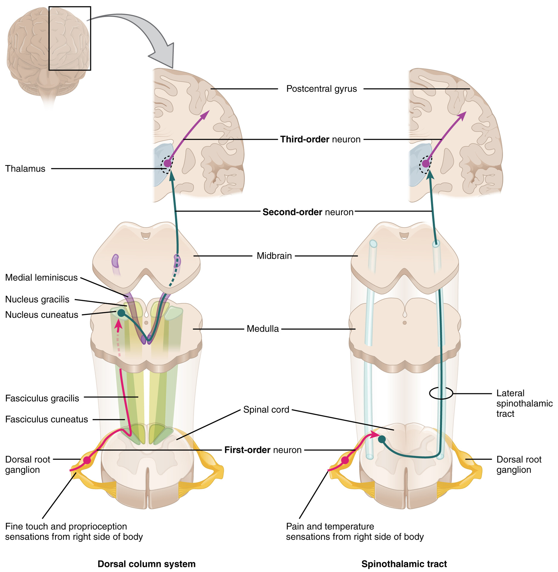
Septomarginal fasciculus and interfascicular fasciculus are little dorsal motor tracts which are barely mentioned in the anatomical literature, and often omitted by medical textbooks. Wycoco, Victor, et al. The cerebellum is important in contributing to the motor system because it compares cerebral motor commands with proprioceptive feedback. The nucleus gracilis is the target of fibers in the fasciculus gracilis, whereas the nucleus cuneatus is the target of fibers in the fasciculus cuneatus. Posture and Gait Gait can either be considered a separate part of the neurological exam or a subtest of the coordination exam that addresses walking and balance.
Sensory motor integration refers to the link between the nerves sensory system and the muscles motor skills and to the process of receiving information through our senses, interpreting it, and organizing it. For the leg, the knee-jerk reflex of the quadriceps is common, as is the ankle reflex for the gastrocnemius and soleus. Watch this Extrapyramidal Controls Other descending connections between the brain and the spinal cord are called the extrapyramidal system. The spinothalamic tracts fibres decussate across the midline at the level of the spinal cord, instead of waiting to get up to the brainstem like the rest of the spinal long white matter structures. What is the path of sensory information? If muscle tone is present, muscle strength is tested by having the patient contract muscles against resistance. Spinal cord tracts: Spinal cord tracts are identified.
The examiner will switch between using the two points and a single point as the stimulus. Testing of the senses begins with examining the regions known as dermatomes that connect to the cortical region where somatosensation is perceived in the postcentral gyrus. One motor neuron connects to multiple muscle fibers within a target muscle. The reason for this is that the dorsal column pathway ascends ipsilateral to the sensation, so it would be damaged the same way as the lateral corticospinal tract. There is a cognitive aspect to remembering how the alphabet goes and how to recite it backwards. After the accident, his personality appeared to change, but he eventually learned to cope with the trauma and lived as a coach driver even after such a traumatic event.
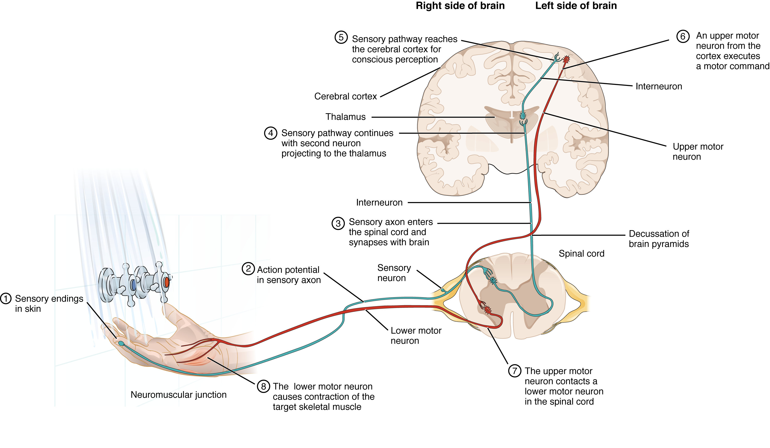
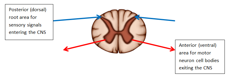
The problem is our brains get overloaded and we begin to become more and more impulsive making it harder and harder to make the sensory pathways work. Typically, spinal nerve systems that connect to the brain are contralateral, in that the right side of the body is connected to the left side of the brain and the left side of the body to the right side of the brain. This weird factoid has relevance: when the central canal is swelled by a syrinx, the medial pain and temperature fibres from the upper limb take the first hit, and sacral and lower limb sensation may be spared, giving rise to a Anterior spinothalamic tract is organised in much the same way i. An overall loss of strength, without laterality, could indicate a global problem with the motor system. When the examiner releases the arm, the patient should be able to stop the increased contraction and keep the arm from moving. All of these motor pathways project to the spinal cord to synapse with motor neurons in the ventral horn of the spinal cord.
What is the difference between sensory and motor pathways?
. Somatic senses inform the nervous system about the external environment, but the response to that is through voluntary muscle movement. For sensations below the neck, the right side of the body is connected to the left side of the brain and the left side of the body to the right side of the brain. Where during their larval stage do they think it belongs? These two systems are similar in that they both begin with dorsal root ganglion cells, as with most general sensory information. As with the previously discussed nerve tracts, the sensory pathways of the trigeminal pathway each involve three successive neurons. This area is responsible for controlling movements of the structures of speech production.
Motor neurons motoneurons carry signals from the central nervous system to the outer parts muscles, skin, glands of your body. The fibres do not decussate with the rest of the big motor bundles in the pyramids -they cross at the level of the spinal nerve. Instead, we experience what can be referred to as a seamless percept. Fibres from here decussate at the level of the brainstem and then travel down the spine to innervate mainly flexor muscles, like the lateral medullary corticospinal tract. Skeletal muscle should have a resting tension representing a slight contraction of the fibers. The assessment of cerebellar function will depend on the normal functioning of other systems addressed in previous sections of the neurological exam.
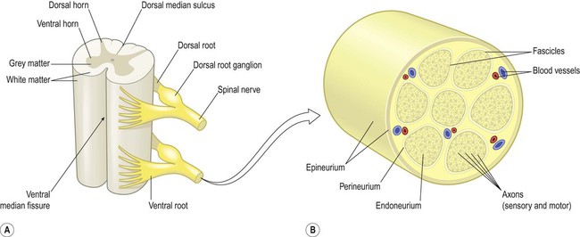
If you're made of money and insist on buying a neuroanatomy textbook at some stage, for whatever reason, make it Snell's Clinical Neuroanatomy by Splittgerber mine is 2018. The point of this is to remove the visual feedback for the movement and force the driver to rely just on proprioceptive information about the movement and position of their fingertip relative to their nose. For children who have sensory difficulties, sensory integration therapy can help sufferers to organize sensory input better. Spinotectal tract runs close to the spinoolivary tract, but unlike the latter it does not cross back to the ipsilateral brain. The medial nuclei serve as a relay for information from the limbic system and basal ganglia to the cerebral cortex. Structure of a Nerve A nerve contains bundles of nerve fibers, either axons or dendrites, surrounded by connective tissue.
Where does sensory information go in the spinal cord? Axons from these second neurons then decussate within the spinal cord and ascend to the brain and enter the thalamus, where each synapses with the third neuron in its respective pathway. The most dramatic example of this is during the overconsumption of alcohol. The upshot of this separation of fibres is not entire clear, from a clinical perspective. However, the somatosensory pathways are divided into two separate systems on the basis of the location of the receptor neurons. Interneurons connect various neurons within the brain and spinal cord. The motor pathways are called descending pathways or descending tracts, because they are traveling south, down the spinal cord, away from the brain.
Ataxia is often the result of exposure to exogenous substances, focal lesions, or a genetic disorder. The most common superficial reflex in the neurological exam is the plantar reflex that tests for the Babinski sign on the basis of the extension or flexion of the toes at the plantar surface of the foot. The tectospinal tract projects from the midbrain to the spinal cord and is important for postural movements that are driven by the superior colliculus. Specifically, the effects of spinal cord transection. The spinothalamic tract travels to the spine, from the thalamus. Conversely, the axons of the corticospinal tract are largely contralateral, meaning that they cross the midline of the brain stem or spinal cord and synapse on the opposite side of the body.