Structure of human eye. Structure of human eye 2022-12-23

Structure of human eye Rating: 6,6/10 1272 reviews

The human eye is a complex and intricate organ that plays a vital role in our ability to see and perceive the world around us. The structure of the eye is designed to allow light to enter and be focused onto the retina, which is the light-sensitive layer at the back of the eye. The retina is responsible for converting light into electrical signals that the brain can interpret as images.

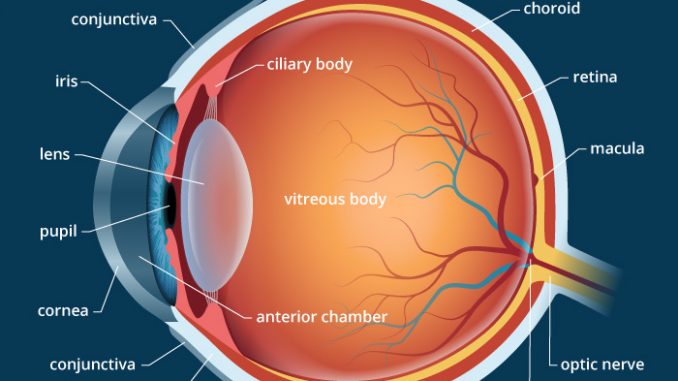
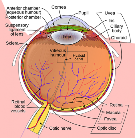
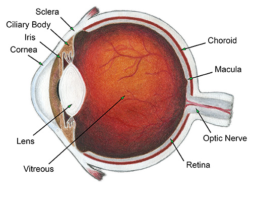
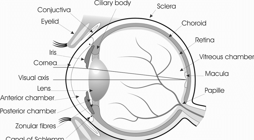
The eye is divided into two main parts: the front part, which is called the anterior chamber, and the back part, which is called the posterior chamber. The front part of the eye is made up of the cornea, the iris, and the lens. The cornea is the transparent outer layer that covers the front of the eye and helps to protect it from dirt, dust, and other foreign objects. It is also responsible for bending the light that enters the eye, which helps to focus it onto the retina.

The iris is the colored part of the eye that surrounds the pupil, which is the small opening in the center of the iris. The size of the pupil can be adjusted to control the amount of light that enters the eye. This is important because too much light can damage the retina, while too little light can make it difficult to see.

The lens is a transparent structure located behind the iris that helps to focus light onto the retina. The lens is held in place by the ciliary muscles, which can change the shape of the lens to help focus on objects at different distances.

The posterior chamber of the eye is made up of the vitreous humor, the retina, and the optic nerve. The vitreous humor is a clear, gel-like substance that fills the space between the lens and the retina. It helps to maintain the shape of the eye and provides a clear path for light to reach the retina.

The retina is a thin layer of cells located at the back of the eye that is responsible for converting light into electrical signals. The retina is made up of several different types of cells, including rods and cones, which are specialized cells that are sensitive to light. The rods are responsible for detecting light and dark, while the cones are responsible for detecting colors.

The optic nerve is a bundle of nerve fibers that carries the electrical signals from the retina to the brain. The optic nerve is located at the back of the eye and is responsible for transmitting the information that the retina has collected to the brain, which then interprets it as an image.

In conclusion, the structure of the human eye is a complex and intricate system that is designed to allow us to see and perceive the world around us. The various parts of the eye work together to allow light to enter the eye, be focused onto the retina, and be converted into electrical signals that the brain can interpret as images.

Structure of human eye (video)

structure of human eye

This is the see-through part of the eyeball. The sphincter pupillae make the pupil contract, whereas the dilator pupillae enlarges the pupil. These nerves connect the brain with the Eye in a different form. This provides you with a sense of vision. When we have to continuously gaze at an object which is moving in a small amount, then this movement helps us. The ciliary muscle changes the shape of the eye lens to help you see clearly.

Next

Eye Anatomy

structure of human eye

Vitreous means glassy, hence the name, because the vitreous humour is very clear so that light can pass through it. Behind the iris, the posterior chamber of the eye starts. It is a bundle of more than one million nerve fibers that forms just behind the retina, connecting it to the brain. Ciliary Muscle The ciliary muscle is situated behind the cornea in a ring-shaped figure present inside the eye. Around 130 million cells are present here. Then it decodes the information to the message or commands. Tiny particles that may be on our eye like specks of dust get washed into the corner of our eyes next to our nose.

Next

Iris of the Eye (Anatomy, Functions & Associated Conditions)

structure of human eye

The eye receives direct oxygen through the aqueous humor. These muscles are both controlled voluntarily and involuntarily to track objects and correct for simultaneous head movements. Occupational and Environmental Medicine. The aqueous humor and the vitreous body are transparent so that there is barely any measurable refraction of single photons. No vision is possible in the blind spot. These cases can develop. Evolution has advanced the eye in such a way that it uses as little energy as possible.


Next

Structure Of Human Eye : Lens to see the world

structure of human eye

It is enclosed in a transparent capsule. These impulses are sent to the optic disk on the retina where they get transferred by a further set of electrical impulses along the optic nerve. As a result, both eyes are unable to look at the same spot at the same time. The human eye is a very sensitive and delicate organ suspended in the eye socket which protects it from injuries. The lens works the same as the camera lens.

Next

Anatomy and Physiology of Human Eye

structure of human eye

This dome-shaped cornea is a transparent dome that will help the light to pass through them up to the lens,resulting in the formation of the image at the back of the eye,that is the retina. Any object you see , the light rays first come through the cornea. Scientists believe that in the ancient era when humans only lived in the caves, they mostly use the Cone Cell for vision. The Optic Disk identifies the start of the Optic Nerves where messages from Cone and Rod cells start from the eye through nerve fibres to the optic centre of the brain. Without it, the shape of the eyes makes gets misshaped due to injury.


Next

Structure of human eye

structure of human eye

Blink frequency is defined as the number of blinks per minute and it is associated with eye irritation. Optical Devices in Ophthalmology and Optometry: Technology, Design Principles and Clinical Applications. This condition majorly occurs due to the wrong or improper connectivity of the nerve connecting the eye to the brain. This is a very smooth movement rather than other movements in the Eye. This is called a Near Response. This area has a high concentration of Cone cells.

Next

Structure and Functions of Human Eye with labelled Diagram

structure of human eye

AMD affects the sight and with it, the power to ascertain fine details. The quality of your farsightedness defines your focusing capability. The cornea itself is composed of five layers, including three cellular layers and two non-cellular layers. The light information is bundled in named receptive fields, enlarged, and transmitted to the brain via the visual pathway. Your doctor may prescribe contact lenses if your iris problem causes poor vision.


Next

Human Eye

structure of human eye

Structure of Human Eye A human eye is roughly 2. The ciliary muscles has to push the lens to increase its curvature. It is the region where the Retina gets connected with the Optic Nerves. Eyelids are attached to the eyelashes. It is clear and slightly convex.

Next

Structure and working of human eye

structure of human eye

Inside the wall of the eyes, the retina is present all over the wall. Having two eyes allows the brain to determine the depth and distance of an object, called stereovision, and gives the sense of three-dimensionality to the vision. This focuses the light rays falling onto it on the retina causing the formation of the image. It consists of two types of nerve cells viz. How The combination of Lens And Eye Works The lens is an important part of the Spherical human eye. The lens forms an inverted real image on Retina.

Next