Temporal bone function. Temporalis: Origin, insertion, innervation, function 2022-12-18

Temporal bone function Rating: 4,9/10 404 reviews

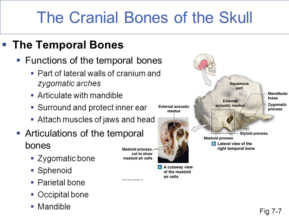
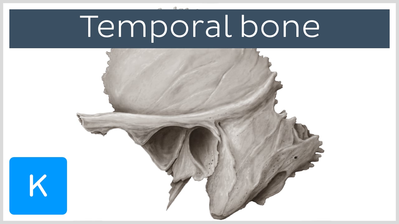
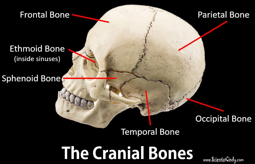
The temporal bones are a pair of bones located on either side of the skull, behind the ears. These bones have several important functions in the body.

First and foremost, the temporal bones serve as protective structures for the brain. They contain the auditory tubes, which allow for the passage of sound waves to the inner ear, and the auditory ossicles, which are small bones that transmit sound waves from the eardrum to the inner ear. The temporal bones also house the structures that are responsible for balance, including the vestibular system and the vestibulocochlear nerve.

In addition to their role in hearing and balance, the temporal bones also serve as attachment points for several muscles. These muscles, including the temporalis and masseter muscles, are responsible for chewing and facial expressions.

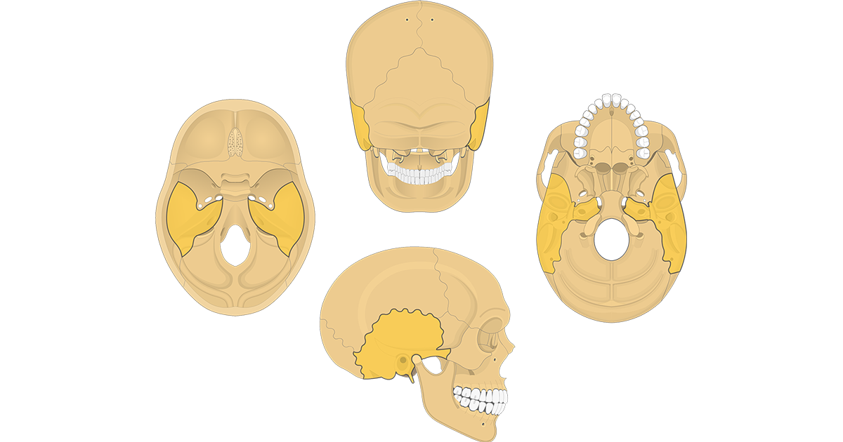
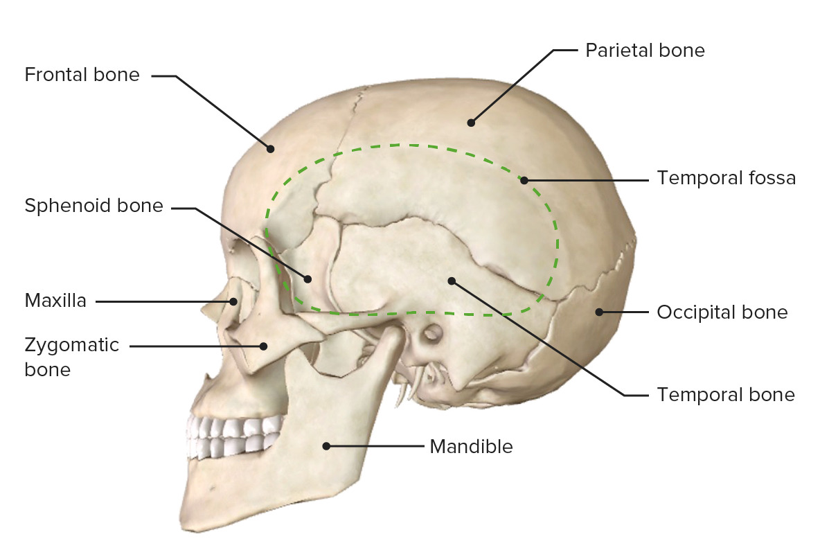
The temporal bones also contain the zygomatic arch, which is a bony arch that spans across the cheek and is important in the movement of the lower jaw. The zygomatic arch is formed by the zygomatic process of the temporal bone and the zygomatic process of the cheekbone.

Finally, the temporal bones contain several important blood vessels and nerves, including the internal carotid artery and the middle meningeal artery, which provide blood to the brain, and the trigeminal nerve, which is responsible for sensation in the face.

In summary, the temporal bones are essential for several important functions in the body, including hearing, balance, facial expression, and the supply of blood and nerves to the brain. They play a vital role in the overall function and health of the body.

Temporal Bone

temporal bone function

. Facial nerve decompression The tympanic and descending segments of the FN can be decompressed in the temporal bone lab Figure 6A and B. Careful bone reading en face, high power magnification is the key to identifying the chorda. Some of the clinical signs that indicate its fracture are hemorrhagic otorrhea or blood leakage through the ear, vertigo, abnormal movement of the eyes, ringing in the ears or tinnitus and Battle's sign which is the hematoma over the mastoid process, among others. The geniculate ganglion is found medial to the STR Figure 5D. The UC Irvine Head and Neck Cancer team includes two neurotologists Drs.

Next

Infections of the Temporal Bone

temporal bone function

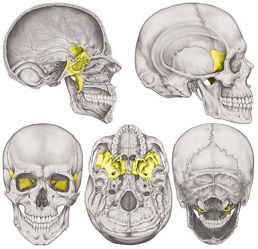
One can feel the zygomatic process by touching just under the eye and moving toward the ear. It forms a bony mass between the sphenoid and occipital bones within the cranial cavity. The contraction of its anterior fibers moves the mandible dorsocranially elevation. Treatment for temporal lobe damage will vary depending on what types of secondary effects the individual is experiencing. Notably, fractures can also lead to leaking of cerebral spinal fluid. Ligaments and joint capsule Synonyms: Superior and inferior synovial membranes of temporomandibular joint, Synovium of temporomandibular joint , The joint capsule originates from the borders of the mandibular fossa, encloses the articular tubercle of temporal bone and inserts at the neck of mandible above the pterygoid fovea, which is a depression on the anterior surface of the neck. It then enters the foramen lacerum, transits the pterygoid canal and enters sphenopalatine fossa through the vidian canal.

Next

Temporomandibular joint (TMJ): Anatomy and function

temporal bone function

The middle fossa plate MFP should be first identified in the squama of the temporal bone from the zygmatic root to the lateral sinus. This maneuver is safe as long as a thin layer bone is kept intact along the IAC. The zygomatic process of the temporal bone articulates with the temporal process of the zygomatic bone of the face, forming a structure called the zygomatic arch. The bone is concave to form the wall for the external acoustic meatus. Then define the course of IAC by drilling from the porus to the fundus. Passing through this opening are the facial nerve CN VII , the labyrinthine artery.

Next

Temporal Lobe Function

temporal bone function

Lobes of the Brain The brain as a whole can be divided into three main parts: the cerebrum, the cerebellum, and the brainstem. Sound enters the ear canal and makes the tiny bones ossicles inside the ear vibrate. A narrow bony projection, called the styloid process, extends downwards and anteriorly from the inferior surface of the tympanic part. Depending on the extent of the tumor, radiation sometimes has to be performed as well. This hollow partially encloses the internal acoustic meatus and the Synonyms: Meckel's groove, Impressio trigemini ossis temporalis The ridge is limited posteriorly by the arcuate eminence, which is raised superiorly by the superior semicircular canal. We hope this article helped you understand the potential outcomes of temporal lobe damage and how to approach recovery.

Next

Temporalis: Origin, insertion, innervation, function

temporal bone function

Temporal Bone Dissection Guide first version, by John K. TemporalBone Where is the Temporal Bone Located The temporal bones are located on the lateral portions of the cranial floor. This tract consists of cells surrounding the fallopian canal Figure 4C and is anatomically variable. The saccule then connects with the endolymphatic sacthrough the vestibular aqueduct. The sensory branch of the V3 that returns to the dura also transits foramen spinosum. We link primary sources — including studies, scientific references, and statistics — within each article and also list them in the resources section at the bottom of our articles.

Next

Temporal bone: Anatomy, parts, sutures and foramina

temporal bone function

During this step, drill superiorly between the MFP and the labyrinthine segment is a safe maneuver to define the entire course of the labynrinthine segment as it is away from the tympanic FN. The surrounding tissue is then dissected away from the EAC. Temporal Bone Diagram The images help with identifying the twenty-two skull bones. Â The inferior compartment allows rotational movements. Anatomy and inflammatory and neoplastic processes.

Next

Temporal bone: function, parts, development, traumas, pathologies

temporal bone function

It is more difficult to control the drill when drilling with the tip and visualization is suboptimal. The groove should extend from the root of the zygoma to the mastoid tip Figure 4A. This division allows its better understanding for its anatomical study. A thin layer of bone should be kept atop. Due to the sunny nature of Southern California, our team has had extensive experience with cancers of the ear.

Next

Temporal Bone Anatomy (Cadaveric Dissection)

temporal bone function

The temporal bone location is at the sides and base of the skull. Doctors also may test the person's hearing and whether the face is paralyzed. If there is dehiscence, the bony wall is usually thin enough to be removed without drilling Per Dr. The posterior EAC wall is thinned using long steady strokes that parallel the descending facial nerve. Let's Play Doctor: The brain is divided into several lobes frontal, occipital, parietal, and temporal. Its outer surface is roughened by muscular attachments. Its upper face is concave and constitutes the posterior wall of the internal auditory canal.

Next

Temporal bone: anatomy, function and injuries

temporal bone function

The bone between the descending FN and the sigmoid sinus is then removed to expose the jugular bulb Figure 11B. The vestibulocochlear nerve terminates in the temporal bone. The EAC wall should be thinned until the cortical plate is identified. If doctors believe After a trauma, the fluid that surrounds the brain, called cerebrospinal fluid, can leak from the ears or nose. Extended Facial Recess After posterior tympanotomy, the chorda is transected along the FN.

Next