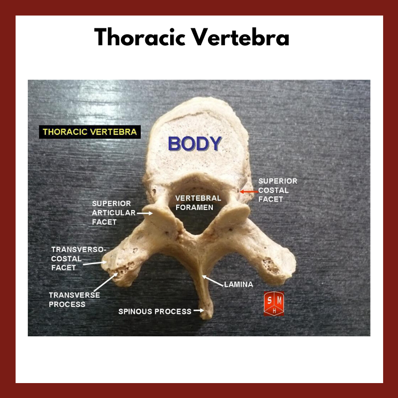
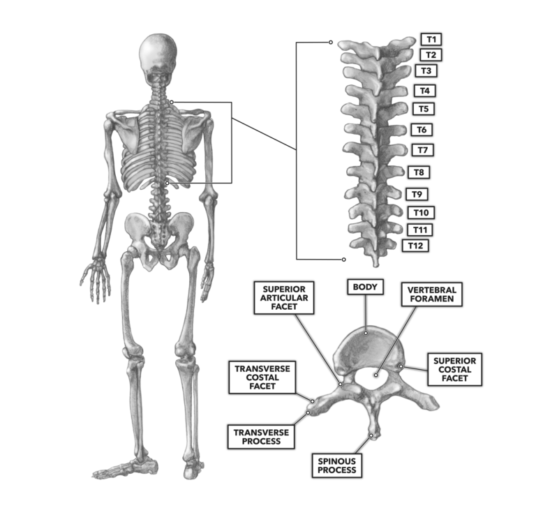
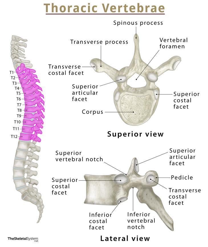
The thoracic vertebrae are a group of twelve vertebrae located in the upper part of the spine, between the cervical vertebrae in the neck and the lumbar vertebrae in the lower back. These vertebrae form a critical part of the human skeleton, providing support and stability for the body while also allowing for a range of movements.
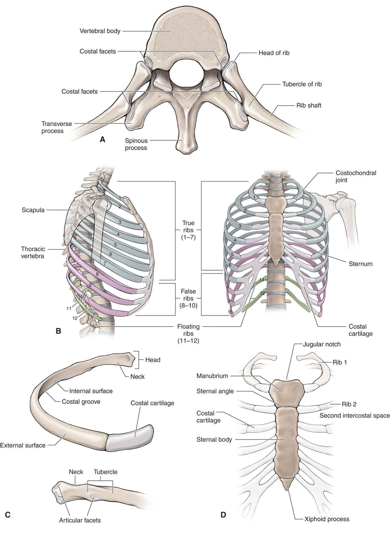
One of the key features of the thoracic vertebrae is their connection to the ribcage. Each thoracic vertebra has a pair of ribs attached to it, with the first rib attaching to the first thoracic vertebra and the twelfth rib attaching to the twelfth thoracic vertebra. The ribs help to protect the vital organs located within the thoracic cavity, including the heart, lungs, and other major blood vessels.
In addition to their role in supporting and protecting the body, the thoracic vertebrae also play a crucial role in enabling movement. The thoracic vertebrae are connected to one another by intervertebral discs and various ligaments, which allow for a range of movements such as bending, twisting, and rotation. These movements are essential for activities such as reaching, lifting, and throwing.
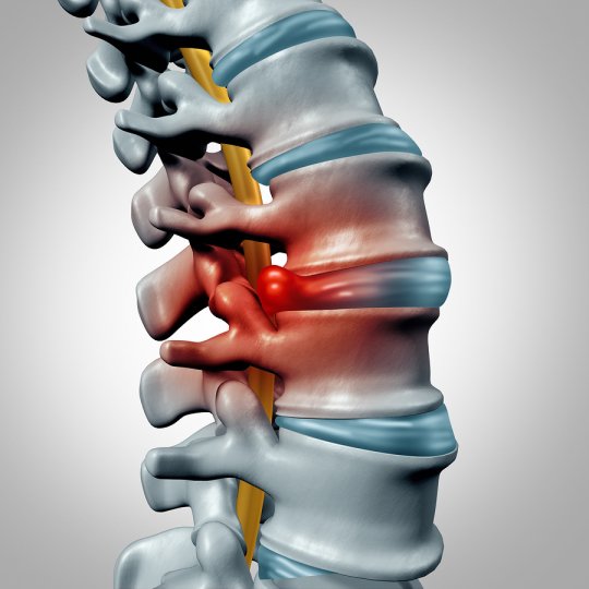
Despite their importance, the thoracic vertebrae are also prone to injury and disease. Osteoporosis, a condition characterized by the loss of bone density, is a common cause of thoracic vertebral fractures. In addition, thoracic spinal stenosis, a condition in which the spinal canal becomes narrowed, can cause pain and discomfort in the upper back.
Overall, the thoracic vertebrae are a vital part of the human skeleton, providing support and stability while also enabling a range of movements. Despite the potential for injury and disease, proper care and maintenance of the thoracic vertebrae can help to prevent these issues and maintain good spinal health.
Vertebral tumor
Last medically reviewed on February 26, 2015. But it's usually not known whether such genetic defects are inherited or simply develop over time. The transverse processes are long, and the upper vertebral notches are deeper than those of the other thoracic vertebrae. The thoracic spine is comprised of 12 vertebrae labeled T1 through T12. American Association of Neurological Surgeons. X-rays can demonstrate if there is any damage to the bones in your spine. The first step is to understand what might be causing your discomfort.
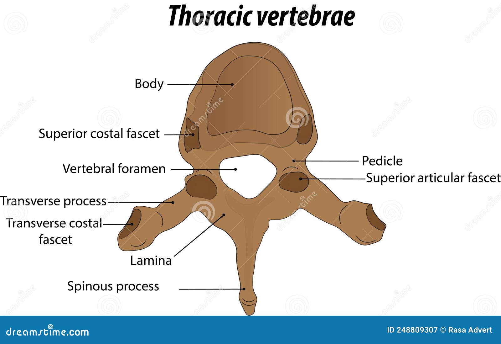
Pedicles Pedicles are cylindrical bony protrusions projecting from the posterolateral surfaces of the vertebral bodies. They alone articulate with the first rib; C7 has no costal facets. This thick, bony front of the vertebra is a rounded heart shape as viewed from above in the thoracic spine region. Symptoms Vertebral tumors can cause different signs and symptoms, especially as tumors grow. These joints are where a vertebra connects, or articulates, with a rib. These transitional forces are due to the change from the rigid thoracic spine to the relatively mobile lumbar spine.
Michaelf- Miami I started using Haka a couple months ago and love my results so far. I am on my second bottle of GLX3. Also, the noted increase in vertebral body size as the spinal column descends is directly related to the increased weight-bearing requirement; further down the column, the greater the proportion of body mass that rests upon it. The motor and sensory functions provided by a thoracic nerve root are determined by its vertebral level. Other possible risk factors include being overweight, smoking, and leading a sedentary lifestyle. History and examination Your doctor will want to know exactly what happened to cause the injury. It extends from the skull to the coccyx and includes the cervical, thoracic, lumbar and sacral regions.
6 Common Thoracic Spine Pain Symptoms and Their Treatments
The top thoracic vertebra, T1, connects with C7 in the cervical spine above while the bottom thoracic vertebra, T12, connects with L1 in the lumbar spine below. Last medically reviewed on February 27, 2015. The second risk factor is age. The first two posterior intercostal arteries branch off of the subclavian artery while the remaining branch off of the thoracic aorta. How are conditions of the thoracic spine diagnosed? Treatment goals include protecting nerve function and restoring alignment and stability of the spine. As it approaches the lumbar vertebrae, the size of the vertebral bodies increases.
Thirty-one pairs of nerves branch out through vertebral openings the neural foramen along your spine. Tumors that begin in the bones of the spine primary tumors are far less common. Scoliosis can range from mild to severe. Vertebrae are the 33 individual, interlocking bones that form your spinal column. The first is obesity.
Last medically reviewed on January 19, 2018. Veritas Health, LLC, 520 Lake Cook Road, Suite 350, Deerfield, IL, 60015. The 12 thoracic vertebrae are labeled T1 through T12, with T1 being closest to the skull and T12 being closest to the tailbone. It helps diagnose bone spurs, osteophytes, bone fusion and bone destruction from infection or tumor. It extends from the base of the skull to the lower back. Thoracic vertebrae nerves Meningeal branches of spinal nerves innervate all thoracic vertebrae.
This projection gradually increases as the column descends before decreasing rapidly from T9-T12. If you have had a recent infection, HIV infection, or drug misuse, you are more likely to experience thoracic spine pain. Your doctor may also request X-rays of your thoracic spine. Traces of similar elevations are found on the transverse processes of the tenth and eleventh thoracic vertebrae. Merck Manual Professional Version. The spinal process is perhaps the most prominent, jutting out like a wing. This bony hole is formed by two adjacent vertebrae, and its size and shape can slightly shift as the vertebrae move.
Thoracic Vertebrae Spine (T1) Images, Model & Pictures
Merck Manual Professional Version. These vertebrae move to allow for a range of motion. Get Mayo Clinic cancer expertise delivered to your inbox. Mayo Clinic, Rochester, Minn. Intervertebral disks allow you to bend and move with ease. Thoracic spine pain is defined as pain in the back that is located between your first thoracic vertebra and your 12th thoracic vertebra. At the T1 through T11 levels, the ventral ramus eventually becomes an intercostal nerve that travels along the same path as the ribs specifically between the innermost and internal intercostal muscles that connect adjacent ribs.
Transverse Processes These are long and thin wing-like structures projecting laterally from the junction between the pedicle from both sides of the vertebrae. Complementary and alternative therapies for cancer. It should not be considered professional advice nor serve as a substitute for medical consultation. As T12 has characteristics of both thoracic and lumbar vertebrae, it is subject to transitional stresses. Other processes include the inferior articular process, the superior articular process, and the transverse process.
Thoracic vertebrae anatomy, function & thoracic vertebrae injury
What is the most common injury to the thoracic spine? There are twelve vertebrae blocks of bone in the thoracic chest region at the back. Anatomy, Back, Vertebral Column 2. Have Always been on serious meds to improve quality of life. A tightened space can cause your spinal cord or nerves to become irritated, compressed or pinched. A person who sustains an injury in this area will most likely experience limited or complete loss of use of the muscles in the lower abdomen, buttocks, legs, and feet. That said, some standard treatments are Thoracic Spine Pain Research How many school lockers are there in the United States? Talk to your healthcare provider about what kind of shoes are best for you and if you should use orthotics or inserts. These main arteries branch out into the periosteal and equatorial arteries, which in turn branch into anterior and posterior canal branches.