The upper extremity, also known as the arm, contains a complex network of veins that play a crucial role in the circulatory system. These veins function to transport blood from the arms back to the heart and lungs to be oxygenated.
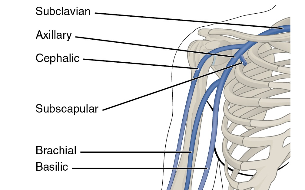
There are two main types of veins in the upper extremity: superficial veins and deep veins. Superficial veins are located close to the surface of the skin and can be easily seen and felt. These veins include the cephalic vein, which runs along the lateral aspect of the arm and wrist, and the basilic vein, which runs along the medial aspect of the arm.
Deep veins, on the other hand, are located deeper within the tissue and are not visible from the surface. The deep veins of the upper extremity include the axillary vein, which runs through the armpit and connects to the subclavian vein, and the brachial vein, which runs along the inside of the arm and connects to the axillary vein.
Both the superficial and deep veins of the upper extremity are important for maintaining proper blood flow and circulation. However, the deep veins are especially vital as they are responsible for carrying the majority of the blood from the upper extremity back to the heart.
The upper extremity veins also have a number of important functions beyond just transporting blood. They help to regulate blood pressure and can act as a reservoir for excess blood during times of stress or physical activity. In addition, the veins of the upper extremity are essential for the administration of intravenous fluids and medications.
Overall, the anatomy of the upper extremity veins is complex and plays a crucial role in the proper functioning of the circulatory system. Proper care and attention to these veins is essential for maintaining good health and preventing problems such as vein damage or thrombosis.
Primary (spontaneous) upper extremity deep vein thrombosis
This is usually for the purpose of providing intravenous therapy e. These devices are inserted in upper extremity veins and are long enough to reach central veins in the chest. The cephalic vein arises from the radial aspect of the dorsal venous network within the Similarly, the basilic vein arises from the dorsal venous network of the hand but from the ulnar aspect, ascending posteromedially within the forearm. The intervertebral veins vv. Therefore, they are absent in the palm fist area , along the ulnar border of the forearm supporting border , and in the back of the arm and trapezius region resting surface.
Lower extremity veins also tend to have more valves, which facilitate the pumping action of muscular contractions. Main superficial veins of the upper limb include the dorsal venous network, and the cephalic and basilic veins. They then unite to form a single trunk, which runs up on the medial side of the artery and ends in the corresponding innominate vein. Joffe HV, Goldhaber SZ. Its ease of access, fixed position and superficial position make the median cubital vein a good site for venepuncture in many individuals. That said, if dialysis is not anticipated for many months, creation of a very distal fistula using a small diameter vein may be attempted before using a more proximal site. There is variability in how the cephalic vein crosses the elbow, and veins in this location are often damaged by prior phlebotomies.
The external jugular vein drains blood primarily from the scalp and face. The veins of the upper extremity are divided into two sets, superficial and deep; the two sets anastomose frequently with each other. Equipment setup Use a mid frequency probe 5-8MHZ. In this article, we shall look at the anatomy of the upper limb veins — their anatomical course, structure, and their clinical relevance. As the diaphragm descends into the abdomen, intra-abdominal pressure increases and thus venous return from the lower extremities decreases during inspiration, resulting in a 180-degree phase shift between flow cycles in the upper and lower extremities Figure 6. The superficial lymph nodes lie along the veins and the deep lymph nodes along the arteries. In order for an upper extremity venous thrombus to be considered a DVT the clot has to seen within the internal jugular IJV , subclavian, axillary or brachial veins.
They end in the intervertebral veins. Meaning they both originate and insert within the hand. Anticoagulation is recommended for three months. In operations for removal of the breast in carcinoma , the axillary lymph nodes are removed, and it sometimes becomes necessary to remove a segment of the axillary vein. All the necessary information on the major veins of the upper limbs with a detailed explanation of the veins of the arm is included in this article. Examine the deep veins from the neck and brachiocephalic vein to the hand. The dorsal venous network is formed just superficial to the dorsal metacarpus and receives venous blood from the dorsal metacarpal veins, along with dorsal digital veins of the first and fourth digits.
For these reasons, vein mapping is best done after the patient has been in a warm room for at least thirty minutes and when well hydrated. This vein, at its commencement, receives the right vertebral vein; and, lower down, the right internal mammary and right inferior thyroid veins, and sometimes the vein from the first intercostal space. It can be subdivided into the superficial system and the deep system. Deep veins of the forearm include the radial and ulnar veins as well as the anterior and posterior interosseous veins. Ann Intern Med 2014; 160:451.
Occasionally, the median antebrachial vein splits in two and drains into both the basilic and cephalic veins. On the other hand, the arteries accompany the deep veins. Cephalic Vein The cephalic vein also arises from the dorsal venous network of the hand. The two are managed differently as the former is a local and generally benign process whereas the later may cause extremity symptoms or embolization. The Veins of the Upper Extremity and Thorax F IG. The digits are supported with three consecutively attached bones called the proximal, middle and distal When it comes to the muscles, they are called the intrinsic muscles of the hand. Then, these courses proximally continue as venae comitantes of radial and ulnar arteries; which further join to form the brachial veins.
It then runs upward along the medial border of the Biceps brachii, perforates the deep fascia a little below the middle of the arm, and, ascending on the medial side of the brachial artery to the lower border of the Teres major, is continued onward as the axillary vein. Dorsal digital veins extend along the sides of the dorsal surface of the phalanges of the hands and unite at the base of the proximal phalanges to form three dorsal metacarpal veins deep veins. It is usually provided with a pair of valves, which are situated about 2. Baarslag HJ, van Beek EJ, Koopman MM, Reekers JA. Upper extremity venous flow is also affected more by the cardiac cycle and is more pulsatile than lower extremity venous flow. Main deep veins of the upper limb include the superficial and deep venous palmar arches, the radial and ulnar veins, anterior and posterior interosseous veins, the brachial veins and the axillary vein.
A communicating branch often connects the dorsal venous network to the cephalic vein around the center of the forearm. Anticoagulation may be considered in non-ancer patients for progression with significant extremity symptoms. Any of these structures may be injured due to repetitive motion such as pitching a baseball or abnormal anatomy. It then crosses the forearm anteriorly, traverses the antecubital fossa, and continues up the anterior upper arm to the deltopectoral grove at the shoulder where it then terminates by joining the axillary vein. Deep vein thrombosis, which tends to initiate in relatively stagnant flow areas behind valve cusps, is more common in lower veins due to the greater number of valves. In: Nathnagel Handbuch der Pathologie und Therapie, Anonymous Wein, Holder 1884. A comprehensive review of Paget-Schroetter syndrome.