White fish mitosis. Stages of Mitosis in the Blastula of a Whitefish 2022-12-25

White fish mitosis Rating: 5,6/10 1577 reviews

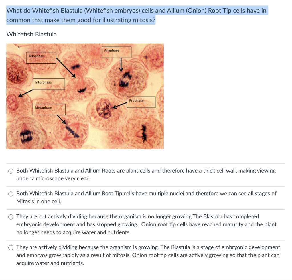
Mitosis is the process by which cells divide and replicate their genetic material in order to produce two identical daughter cells. This process occurs in all organisms and is essential for growth, development, and repair. White fish, like all other organisms, undergo mitosis in order to produce new cells and maintain the integrity of their genetic information.

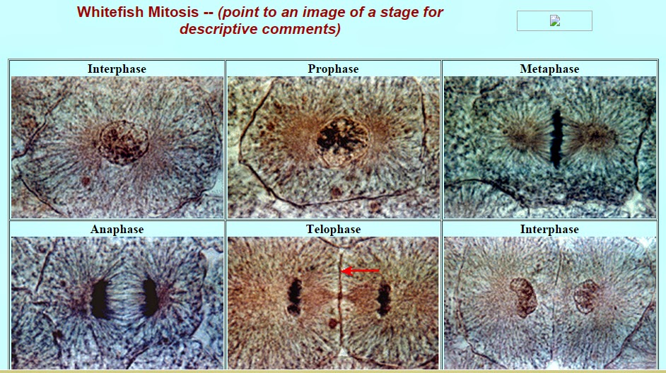
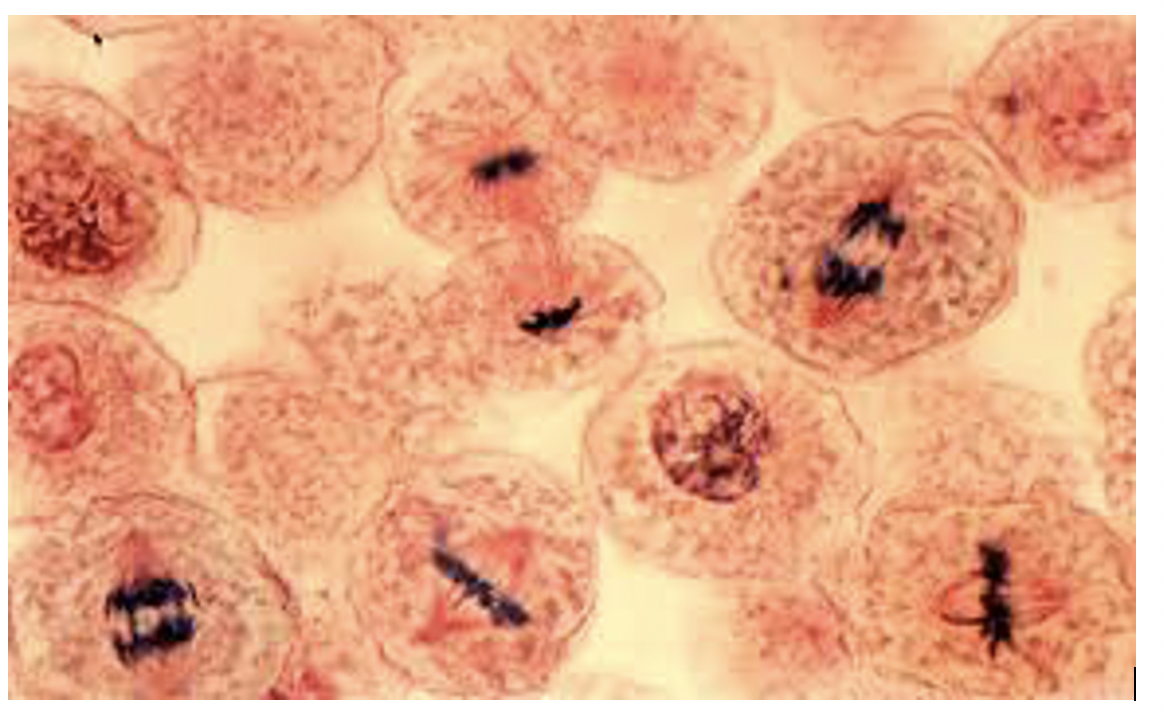
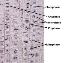
The process of mitosis can be broken down into four main phases: prophase, metaphase, anaphase, and telophase. During prophase, the first phase of mitosis, the nucleolus and nuclear envelope disappear, and the chromatids, which are copies of the chromosomes, become visible.

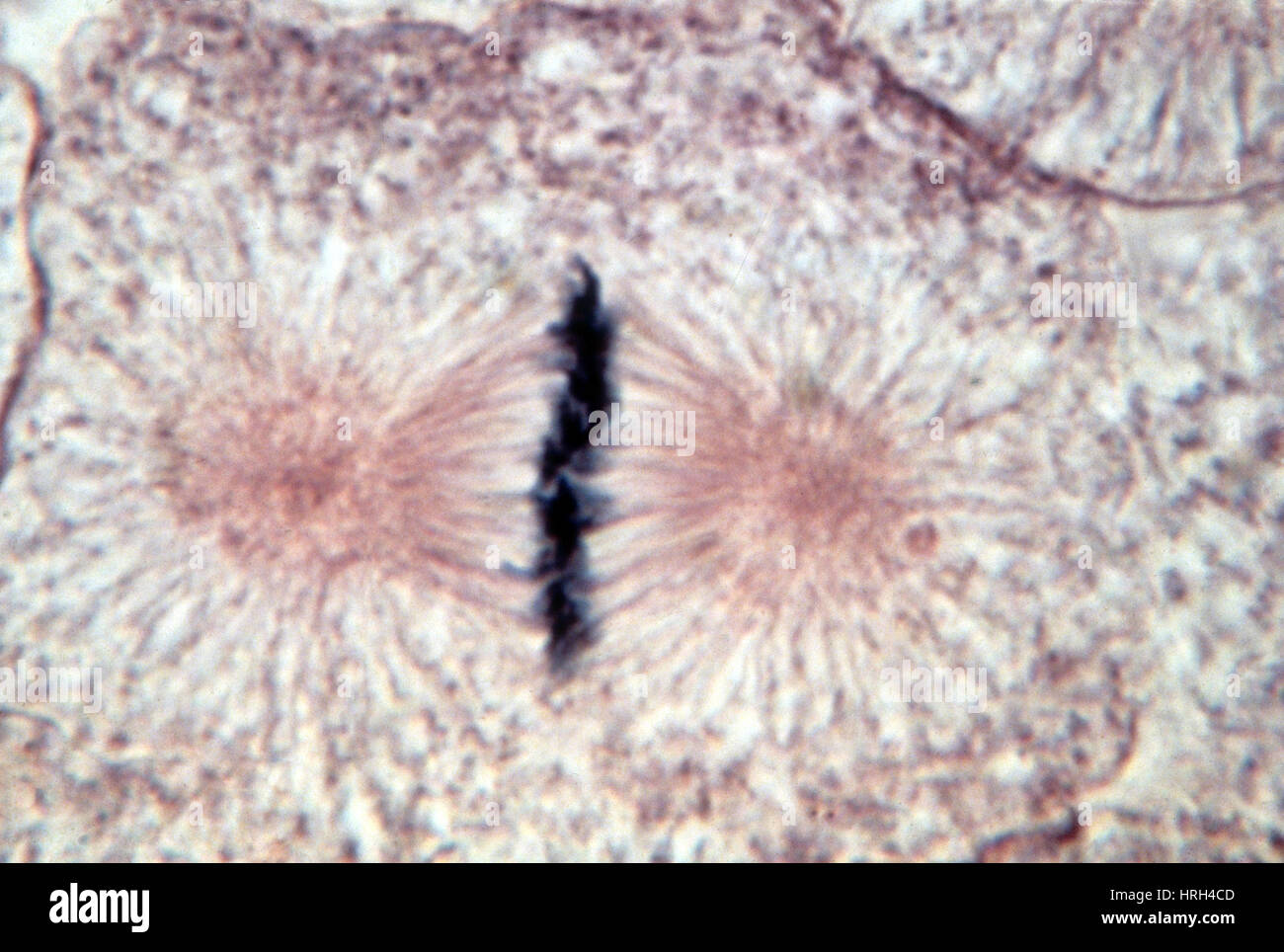
During metaphase, the second phase of mitosis, the chromatids line up at the center of the cell, forming the metaphase plate. This is done through the action of microtubules, which are long, thin structures that help to organize the chromosomes.

During anaphase, the third phase of mitosis, the chromatids are separated and pulled towards opposite poles of the cell. This is done through the action of the mitotic spindle, a structure made up of microtubules that helps to pull the chromosomes apart.

Finally, during telophase, the fourth and final phase of mitosis, two new nuclei are formed, and a cell plate is formed between them, eventually becoming the cell wall. This marks the end of mitosis, and the cell is now considered two daughter cells.

White fish, like all other organisms, undergo mitosis in order to produce new cells and maintain the integrity of their genetic information. This process is essential for the growth and development of white fish, and it ensures that they are able to maintain their genetic diversity, which is important for their survival in the wild. Understanding the process of mitosis is crucial for scientists and biologists who are studying the growth and development of white fish, as well as other organisms.

Slide, Whitefish Mitosis, sec.

white fish mitosis

Note: It is not visually possible to separate the stages of interphase from each other, but the mitotic stages are readily identifiable. Human chromosomes on the other hand, are are not clearly visible at higher power magnification. For each view, identify the stage of mitosis. Each picture contains at least one cell at each stage of mitosis and some stages are represented by multiple cells. Anaphase A whitefish blastula cell in anaphase During anaphase the mitotic spindle apparatus pulls the sister chromatids of each chromosome apart by attaching to each centromere and then pull the chromatids to each pole of the cell. The nucleus is indicated by a blue spot inside of the cytoplasm. Step 2: Student-Led Planning: Look at Figure 13.

Next

Interphase Whitefish Blastula Cell

white fish mitosis

Step 5: Critical Analysis: Are the predictions you made supported by your data observations and calculations? But, the chromatin of the nucleus B appears prominent after staining. Preparation includes chromosome replication, replication of cellular organelles, including centrioles, and the synthesis of microtublule units. Mitosis is also vital for development. Our products are durable, reliable, and affordable to take you from the field to the lab to the kitchen. Test your hypothesis : Test your hypothesis by doing the following: 1. Many single-celled organisms depend on mitosis as their sole or primary way of asexual reproduction.

Next

Onion Root Tip and Whitefish Blastula

white fish mitosis

Mitosis: Onion Root Tip Appearance Under Microscope INTERPHASE Square-like cell membrane with a very solid circle cytoplasm. Why or why not? During prophase, the chromosomes coil up, and sister chromatids become visible under a microscope. The blastula is an early stage of embryo development and represents a period in the organism's life when most of the cells are constantly dividing. Note: There are four major phases of mitosis, plus prometaphase, which is a transition phase between prophase and metaphase. Cancer is the uncontrolled growth and mitotic division of cells which can be caused by mutations in genes. Moreover, the dividing cell have very large and easily seen chromosomes, so its easy to find lots of cells in each stage of mitosis. Other fibers radiate out from the centrioles to create the aster.


Next

Stages of Mitosis in the Blastula of a Whitefish

white fish mitosis

Mitosis is a controlled process and loss of control can lead to cancerous cells. Before proceeding, state your hypothesis. The chromosomes at this stage are dispersed and not visible using a light microscope. Each pair of sister chromatids has a protein structure, called a kinetochore, which becomes attached to spindle fibers. If 100 cells are examined, the number of cells in each identifiable cell- cycle stage will give an estimate of the time it takes for the cell to complete that stage.

Next

Prophase Whitefish Blastula Cell

white fish mitosis

During anaphase, the sister chromatids separate and move to opposite poles of the cell. Activity 1: Observe the Stages of Mitosis in the Blastula of a Whitefish A fundamental property of somatic nonreproductive or body cells of multicellular organisms is mitosis which basically provides new cells for growth and regeneration or replacement of dying and dead cells of the living body. You may want to tape the slide in place on your table. Cancer cells are not healthy cells. Retinoblastoma is the most common primary childhood cancer which often stems from a genetic background.

Next

Whitefish Mitosis

white fish mitosis

G2 Phase The cell prepares the enzymes and machinery for mitosis. Label the prophase diagram: 1. In eukaryotes, all chromosomes must be duplicated prior to mitosis and cytokinesis to ensure each new daughter cell has the full complement of genetic information. During late prophase, the nucleoli disappear and the mitotic spindle apparatus assembles. View 1 View 2 View 3 View 4 View 5 Whitefish Anaphase Metaphase Prophase Telophase Onion Anaphase Metaphase Prophase Interphase Telophase What are the similarities between the two types of cells onion and whitefish? G1 is the phase of growth when the cell is accumulating resources to live and grow. Some chemotherapy drugs, including taxanes such as Taxol from the Yew tree and alkaloids from the Vinca plant, interfere with mitosis by binding to microtubules and preventing spindle fibers from separating sister chromatids, thus leading to cell death. The nuclear membrane surrounding the chromosomes also disappears.


Next

Whitefish (Coregonus) slide, mitosis, section

white fish mitosis

Little to no chromosomes are seen inside the cytoplasm during interphase. Click on the Whitefish embryo and the onion root tip. After attaining a certain size and having amassed enough raw materials, a checkpoint is reached where the cell uses biochemical markers to decide if the next phase should be entered. Label the Interphase diagram: 1. The chromosomes at the end of G1 consist of a single chromatid. If conditions are favorable, the cell continues on to mitosis.

Next

Whitefish Mitosis

white fish mitosis

Science can be messy. Telophase A whitefish blastula cell in telophase + cytokinesis Chromosomes begin to disperse. Whitefish Mitosis Practice The photomicrographs below show sections of whitefish blastula. The mitotic spindle will consist of microtubules that extend from pole to pole. Interphase Whitefish blastula cells in interphase The three phases of Interphase G1 Phase Growth: the cell grows in size and carry out their normal day to day activities. Certain DNA sequences on the ends of the chromosomes called telomeres become shorter during every mitotic cycle of somatic cells, a regulatory mechanism, that contributes to the number of mitotic cycles.

Next

Mitosis Whitefish Blastula Flashcards

white fish mitosis

Exposure of tumor in the right eye by white light reflection. The spindle fibers pull the sister chromatids apart, toward opposite ends of the cell. Distinct chromosomes are not visible to the microscope at this time. They're not happy until you are. Mitosis is part of the cell cycle See Figure 13.

Next

Mitosis in Whitefish Onion Roots

white fish mitosis

The blastula is an early stage of embryo development, and represents a period in the organism's life when most of the cells are dividing consistently. Healthy cells die through programmed cell death or apoptosis after a number of generations of cell divisions. . This asexual reproduction is distinct from multicellular organisms which undergo meiosis to produce reproductive cells sperms or eggs. In your notebook, make a table defining the characteristics of the stages of mitosis, as well as interphase, that you can use to identify each stage under the microscope. When the DNA synthesis is complete, the cell continues on to the second growth phase called G2. METAPHASE Chromosomes which are blue lines under the microscope, are brought to the center by barely visible lines called spindle fibers.

Next