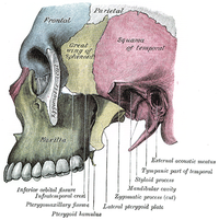
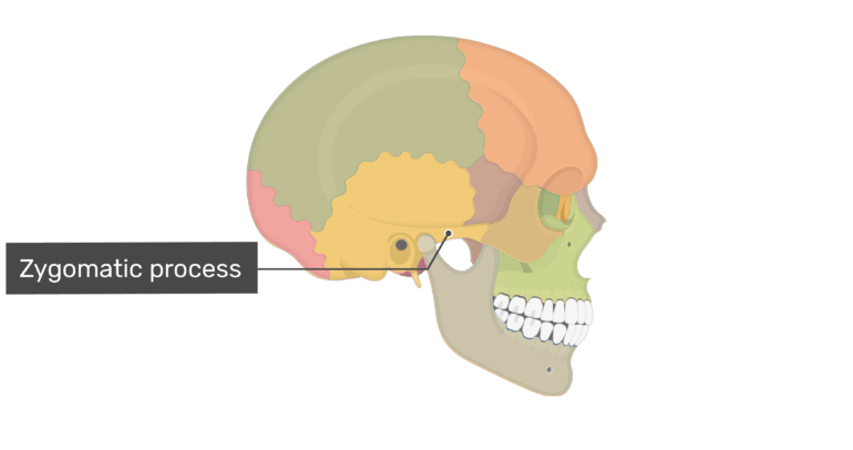
The zygomatic process, also known as the zygomatic bone or cheek bone, is a prominent bony structure located in the face that serves a number of important functions. It is a flat bone located just below the outer corner of the eye and extends outward toward the lateral (side) aspect of the face. The zygomatic bone is formed by the fusion of two bones during fetal development, the zygomatic and temporal bones.
One of the primary functions of the zygomatic bone is to provide support for the facial muscles that are responsible for facial expression. The zygomatic bone is connected to a number of muscles in the face, including the orbicularis oculi, which helps to close the eyelids, and the zygomaticus major, which helps to lift the corners of the mouth and form a smile. The zygomatic bone also helps to protect the eye from injury by acting as a buffer against blows to the side of the face.
In addition to its role in facial expression and protection, the zygomatic bone is also important for the overall shape and appearance of the face. It is a prominent feature of the face and helps to define the cheekbones and give the face a more defined and angular appearance. This is why the zygomatic bone is often a focus of attention in cosmetic procedures such as cheek augmentation, in which the bone is reshaped or augmented to improve the appearance of the face.
The zygomatic bone is also important for the sense of smell. It contains small, thin-walled air spaces called the paranasal sinuses, which are connected to the nasal cavity. These sinuses help to humidify the air that we breathe and also help to reduce the weight of the skull.
In summary, the zygomatic bone is a vital and multifunctional structure in the face that plays a role in facial expression, protection, appearance, and the sense of smell. It is an important structure to consider in a number of medical and cosmetic procedures and is a key feature of the human face.
It is rough at the top and pointed, which helps it articulate with the zygomatic bone. Laterally and dorsally, the frontoethmoid suture is formed by the union of the cribriform plate with the medial surface of the frontal bone. Further exposure of the zygomaticofrontal junction or inferior orbital rim is necessary for severely displaced fractures, which require additional fixation. Like all joints that are incongruous, a fibrocartilaginous disc commonly referred to as a meniscus is interposed to level out the incongruity. Because they are connected to the eye socket and the jawbone, injuries, irritation or disease in these areas can affect the zygomaticus as well. It faces towards the orbit and forms the anterolateral part of its floor and the anterior part of its lateral wall.
Is a fractured cheekbone painful? You will find an orbital part, a temporal surface, a frontal squama, and a nasal part in the frontal bone. You will find alveolar processes in the ventrolateral surface of the maxilla of the dog. You may get help from the below-mentioned dog skull bone diagrams. This both increases the speed of digestion and allows mammals to expand their diet to almost any food they can take a bite out of and chew up. Keyhole defects have been described with handguns and shotguns as well. The external surface of the maxilla bone of a dog has very prominent features, and that is the infraorbital foramen.
You will find a body and four different processes in the maxilla bone of a dog. The caudal recess of the discotemporal compartment is larger than the rostral recess, and the rostral recess of the discomandibular compartment is larger than the caudal recess. The front portion of the bone is thick and jagged to allow for its joining with other bones of the face. You will also find the rostral clinoid process at each side of the caudal end of the preshpenoid bone of the dog. Interparietal and parietal bones of a dog The interparietal bone of the dog skull fuses with the squamous part of the occipital bone before birth and forms the interparietal process. Though not always visible to the naked eye, such fractures can cause internal bleeding and may affect brain function. How do you fix a zygomatic fracture? This crest also continues with the subsequent bone frontal of the skull of a dog.
The much larger masseter muscle which is used for moving the jaw, attaches directly to the zygomatic arch, and the other side to the large part of the mandible. Depending on the severity of the fracture, the zygomatic bone may be monitored through home health and treated with antibiotics to prevent or treat infection. It articulates with the bodies of both presphenoid and basisphenoid bones of a dog. This acts as a partition in the middle of the infratemporal and anterior surfaces. With the origin of the majority of the extraocular rectus muscles in the apex of the orbit, these muscles span the orbit to insert anterior to the globe's equator. There is a nasal crest at the nasal surface of the palatine bone that articulates with the vomer bone in a dog. This kind of a fracture can involve bones of the upper jaw, lower jaw, cheeks, and nose or eye sockets.
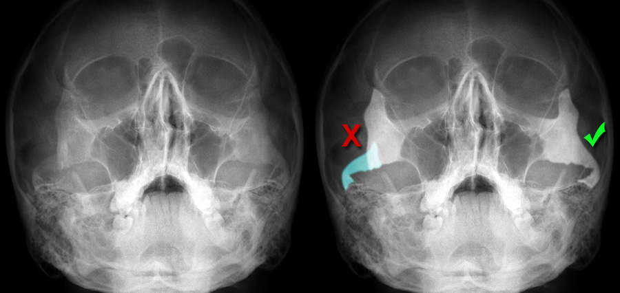
The major arteries in the dog orbit are located along the ventral and ventromedial floor, and consist of the pterygopalatine portion of the maxillary artery with its various branches, including the external ophthalmic orbital artery, the infraorbital artery, the minor palatine artery, and a trunk that gives rise to the major palatine and sphenopalatine arteries. The entrance and exit are contiguous on the bone. The zygomatic bones are more commonly known as the cheekbones. This function is followed by the deepening of the nasolabial groove. The sub-Tenon's space between the fascia bulbi and the sclera is an important surgical plane that permits dissection around the globe with minimal hemorrhage.
Two ligaments support the joint capsules in maintaining joint alignment Figure 101-3. The temporal muscle is then reflected anteriorly through a subperiosteal dissection Fig. This process extends rostrolaterally, overlies the caudal half of the zygomatic bone, and forms the zygomatic arch. What is the left zygomatic arch? Reposition if blood is obtained. Zygomaticus major muscle Musculus zygomaticus major Zygomaticus major is a thin paired buccolabial group of facial muscles along with levator labii superioris alaeque nasi, The main action of zygomaticus major is to pull the angle of the mouth superolaterally.
The globe was retained in 89% of cases and was visual in 78%, suggesting that horses diagnosed with orbital fractures have a favorable prognosis for vision. There is a distinct mastoid process at the petrosal part of the temporal bone. If the zygomatic arch allows for muscle attachment, and reptiles and birds lack these structures, is it impossible for a bird or reptile to have a powerful jaw? This meniscus has an elongated, roundish appearance, is approximately 5 mm thick at the border, and divides the joint into two separate compartments Figure 101-2. Temporal bones of a dog The temporal bone of the dog forms a large part of the ventrolateral wall of the calvaria. The bone must be strong enough, and the shooting really tangential, so that the bone can support the track of the bullet without bursting. To avoid this, people with cracked zygomatic arches are supposed to chew as little as possible during the healing process, which could take months.
Semilunar defects, grooves, gutter wounds, and keyhole defects have been described. Malunion is the most common complication of zygomatic fractures and is the result of improper reduction and fixation, resulting in malocclusion, facial asymmetry, and enophthalmos. Zygomatic Arch Definition The zygomatic arch, cheek bone, or zygoma are all interchangeable terms for the structure in the skull seen indicated by the arrow in the following image. You might identify the following osteological features from the dog mandible. What forms the apex of the skull in dogs? Gray's Anatomy 41tst ed. You can injure these muscles by overuse or trauma.
You will also find a short condyloid canal at the medial side of each condyle. A small artery and vein parallel this nerve. The dorsal surface of the palatine process forms the floor of the ventral nasal meatus. Its anteriormost portion is rough and serves for the articulation with the zygomatic malar process of zygomaticotemporal foramen which transmits the zygomaticotemporal nerve from the orbit to the temporal fossa. Prolonged use of the facial muscles -- such as smiling, talking or chewing a lot during a social event — or physical trauma to the area can cause this condition. This muscle provides most of the motion of the jaw, while the temporalis muscle provides extra tension on the jaw to grind, chew, strip and mash food. This bone articulates with the nasal bone and maxilla rostrally.