B galactosidase molecular weight. Galactosidase, Beta 2022-12-13
B galactosidase molecular weight Rating:
5,4/10
633
reviews
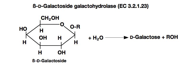
B-galactosidase is a hydrolytic enzyme that catalyzes the breakdown of lactose, a type of sugar found in milk and other dairy products. It is produced by a variety of bacteria, including Escherichia coli, and is commonly used in molecular biology and biotechnology to aid in the digestion and modification of DNA and other biomolecules.
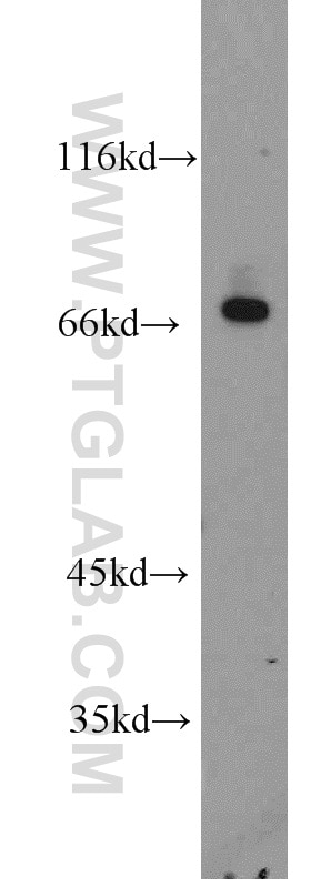
One of the key characteristics of b-galactosidase is its molecular weight, which is a measure of the size and mass of the enzyme. The molecular weight of b-galactosidase varies depending on the specific strain of bacteria it is derived from, but it is typically in the range of 110,000-130,000 daltons. This is relatively large compared to other enzymes, which often have molecular weights in the range of tens of thousands of daltons.
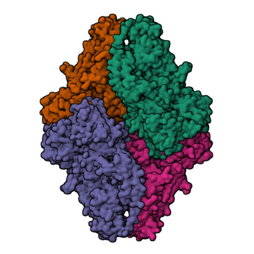
The large size of b-galactosidase is due to the fact that it is composed of multiple subunits, each of which contributes to the overall molecular weight of the enzyme. In the case of E. coli b-galactosidase, there are four subunits, each with a molecular weight of approximately 30,000 daltons. These subunits are held together by non-covalent bonds, which allow them to function as a single unit and perform their catalytic activity.
B-galactosidase has a number of important biological functions, including the digestion of lactose in the gut of mammals and the synthesis of certain biomolecules in bacteria. It has also been widely used in molecular biology and biotechnology as a tool for the analysis and modification of DNA and other biomolecules. For example, b-galactosidase can be used to cleave DNA at specific sites, allowing researchers to manipulate and study the function of specific genes.
In summary, b-galactosidase is a large enzyme with a molecular weight in the range of 110,000-130,000 daltons. It is composed of multiple subunits and plays important roles in both biology and biotechnology. Understanding the molecular structure and properties of b-galactosidase has allowed researchers to develop a range of techniques and applications that rely on this enzyme, including the analysis and manipulation of DNA and other biomolecules.
Galactosidase, Beta
Extinction coefficient of the released product is 33,800 Lmol -1cm -1. Note the increased dpERK signal in lola RNAi guts compared with control. Mean apoptotic cell density values were normalized as a percentage to the wild-type nasal value 100% apoptotic density C. Scale bars, 10 mum. Nuclei are stained with DAPI blue. StrutsRequestWrapper 1c24438a Request attributes: pageContext.
Scale bars, 100 mum. Middle and right panels show higher magnification from the boxed regions. Scale bar, 75 um. After cleavage of the signal peptide, and carbohydrate modifications in the Golgi and lysosomes, the mature enzyme has a native molecular mass of 101kDa. Distribution of the neural crest-derived cells in the brain of dTg mice. Nuclei are stained with DAPI.
Engraftment ratios are noted. Deficiencies in the protein in humans can result in galactosialidosis or Morquio B syndrome. This antibody, raised in rabbit against E. B Mesangial expansion from eAdam17 model. G-H Wing imaginal discs from controls G or expressing Dmp53 H under the control of en-Gal4 ; tub - Gal80 ts and stained to visualize egr -lacZ green or grey and DAPI blue. The alcohols, methanol, ethanol, i-propanol, and n-propanol, at 5% concentration, all increase the rate of o-nitrophenyl, β-D-galactopyranoside cleavage Shifrin and Hunn 1969. Characteristics of β-Galactosidase from E.
Posci et al 1993 Comparison of several new galactosidase as substrates for various β-D-galactosidases. This product was only detected from flies with the intact gypsy-CLEVR transgene. E Immunostaining for PAX8 a secretory cell marker and Ac-TUB a ciliated cell marker in tissue sections of PND35 a and b and PND112 c-f mouse oviducts showing that lacZ + epithelial cells are both secretory and ciliated in nature. The complementary DNA is 1290 bases and encodes a polypeptide of 429 amino acids including a 31 amino acid signal peptide. Notably, some of the α-1- C-alkyl DAB derivatives were also found to have potent human lysosome β-glucosidase inhibitions.
No discernible Mid expression could be seen in an MP2 at 5 hpf, but a faint expression of Mid is seen in MP2 in 6. In addition, the products generated from these processes were highlighted. Galactosylation is the first chemical step in the reaction where Glu461 donates a proton to a glycosidic oxygen, resulting in galactose covalently bonding with Glu537. Merged images are shown in A' and B' green and red. The Journal of Biological Chemistry.
Two D-glucose acts as the acceptor in the second step, transgalactosylation occurs. A shorter Kdm3a protein product result of Cre-induced deletion is indicated DeltaJC, black arrow. Sections were stained for NCCs betagal, green and nuclei DAPI, blue. Right panel shows Kaplan-Meier survival curves. More than 500 mutations have been identified in the gene for α-galactosidase A The Human Gene Mutation Database at the Institute of Medical Genetics in Cardiff, 2011. They achieved the same types of results using a pig endothelial cell line expressing α-galactosidase cDNA. VE-PTP expression in adult kidneys in health and diabetes.
Due to the low pH in this cellular compartment the receptor—ligand complexes dissociate and α-galactosidase A is delivered to lysosomes. This extracellular α-galactosidase A can bind to plasma membrane located mannose-6-phosphate receptors, which mediates its endocytosis and transport to the lysosomes. The presence or absence of an active beta Galactosidase may be detected through addition of artificial chromogenic substrates such as X-gal, fluorescent substrates such as Fluorescein di-beta-D-galactopyranoside FDG , luminescent substrates and others. The arrow shows complete tumor regression, 14 days after Ad-F512 administration. Right panel shows Kaplan-Meier survival curves. In the Pax6 mutant these cells arrowheads in C, D are very scarce and do not contact the pm. Extinction coefficient of the released product is 42,500 Lmol -1cm -1.
N-linked carbohydrate appears at six sites in the glycoprotein dimer, revealing the basis for lysosomal transport via the mannose-6-phosphate receptor Bishop et al. At least 12 MP2s were examined, and all yielded the same patterns of Mid and AJ96 distribution. The anterior end is up, the midline is marked by vertical lines. After treatment of the recombinant enzyme with α-mannosidase, Man-6- P was exposed at the nonreducing end of the glycan moiety, leading to an efficient uptake by fibroblasts derived from patients with Fabry disease. D qRT-PCR showing eiger mRNA levels in wing discs expressing RA CS or dmyc RNAi with en-Gal4.