Coronoid fossa of humerus. Why coronoid fossa of humerus? Explained by FAQ Blog 2023-01-01
Coronoid fossa of humerus Rating:
9,8/10
1227
reviews
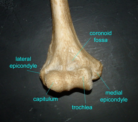
The coronoid fossa is a small depression located on the anterior (front) aspect of the humerus, a long bone in the upper arm. It is named after the coronoid process of the ulna, which is a bony projection located in the forearm. The coronoid process fits into the coronoid fossa when the arm is flexed at the elbow joint.
The primary function of the coronoid fossa is to provide a surface for the coronoid process to articulate with when the arm is flexed. This allows for a smooth and stable movement at the elbow joint, which is essential for activities such as lifting, pushing, and pulling.
The coronoid fossa is also important for the stability of the elbow joint. When the arm is extended, the coronoid process fits snugly into the coronoid fossa, providing a strong connection between the humerus and the ulna. This helps to prevent the elbow from dislocating or moving out of place.
In addition to its role in elbow joint function, the coronoid fossa also serves as an attachment point for several muscles and ligaments. The brachialis muscle, which is responsible for flexing the elbow, inserts into the coronoid process and the adjacent area of the humerus. The ulnar collateral ligament, which helps to stabilize the elbow joint, also attaches to the coronoid fossa.
Overall, the coronoid fossa is a small but important structure that plays a crucial role in the function of the elbow joint. Its unique shape and location allow for smooth and stable movement at the elbow, and it provides attachment points for important muscles and ligaments that help to support and stabilize the joint.
Coronoid Fossa
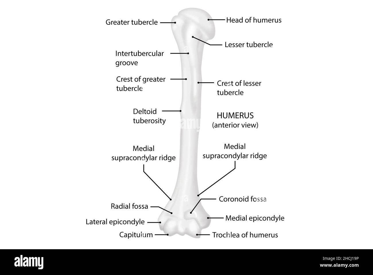
It is directed medially backwards and upwards. Kenhub does not provide medical advice. Clinically oriented anatomy 8th ed. The Lesser Tubercle The lesser tubercle is smaller, anterolaterally placed to the head of the humerus. It articulates with the cupshaped depression on the head of the radius, and is limited to the front and lower part of the bone.
The spiralling nature of the trochlear groove results in the varying transverse axes of the elbow joint. Here it may receive another articulating bone or act to support brain structures. In addition, it serves as the attachment for ligaments and muscles that act on the elbow joint. What articulates with the coronoid fossa? The capitulum of the humerus articulates with the head of the radius. This ridge offers an origin to the lateral head of the triceps brachii.
It is the trochlea of the humerus which sits in the semi-lunar notch of the ulna to form this joint. Nutrient foramen The nutrient foramen of the humerus is located in the anteromedial surface of the humerus. Is the capitulum medial or lateral? The epicondyles, one on either side of the bone, provide attachment for muscles concerned with movements of the forearm and fingers. Anatomy of the Elbow. The mnemonic of the order of appearance of the individual ossification centers is C-R-I-T-O-E: Capitellum, Radial head, Internal medial epicondyle, Trochlea, Olecranon, External lateral epicondyle.
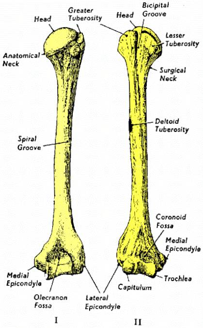
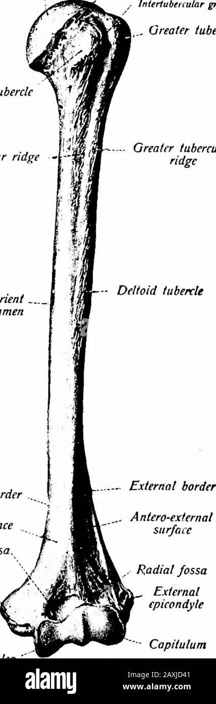
Neck of the Humerus The anatomical neck is the groove that surrounds the articular surface of the head of the humerus. The lower end of the bone presents a small cylindrical head that articulates with the radius at the side and the wrist bones below. The shaft then is connected to the lower part of the humerus which is triangular in shape when viewed in cross-section. The surgical neck however, is a segment present inferior to the humeral head. The lateral portion of the brachialis muscle originates from the distal part of this surface, as well as from the proximal two third of the lateral supracondylar ridge.
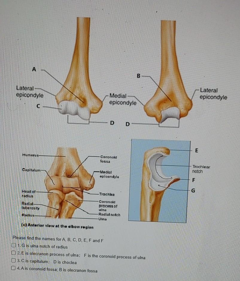
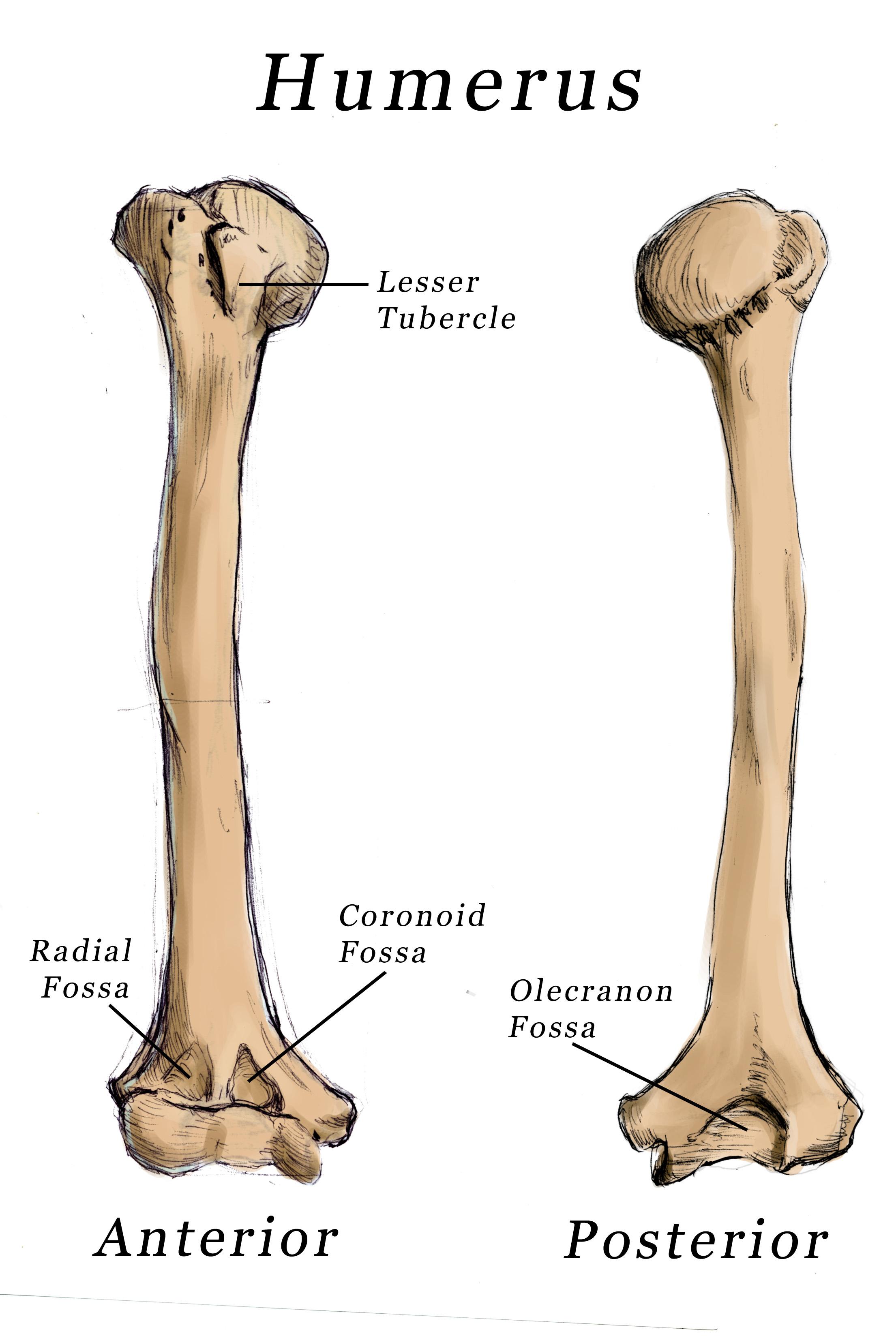
The coronoid fossa is larger than the olecranon fossa and receives the coronoid process of the ulna during maximum flexion of the elbow. Medial Epicondyle The medial epicondyle is the non-articular medial bulge of bone located superior and medial to the trochlea. Deltoid Tuberosity The Deltoid tuberosity is a roughened surface on the lateral surface of the shaft of the Humerus and acts as the site of insertion of deltoideus muscle. Trochlea Trochlea humeri The trochlea has a surface shaped like a pulley and covers the anterior, posterior and inferior surfaces of the medial condyle of the humerus. The radial groove is a shallow groove that interrupts the lateral border in its medial third.
The rami are two vertical processes located on either side of the body; they join the body at the angle of the mandible. What part of the ulna articulates with the humerus? The lateral surface of the coronoid process forms the radial notch for the head of the radius , while the superior surface forms the trochlear notch for the trochlea of the humerus. What is ulna Trochlear notch? It has a smooth proximal surface and is largely covered by the deltoid muscle. The upper end of the ulna presents a large C-shaped notch—the semilunar, or trochlear, notch—which articulates with the trochlea of the humerus upper arm bone to form the elbow joint. During elbow extension, however, the oblique posterior part makes contact with the trochlear notch on the ulna so that this obliquity forces the main axis of the forearm to form a small angle with that of the upper arm. It has 3 surfaces, namely: Anteromedial Surface This is the area between the medial border of the humerus to the line drawn as a continuation of the crest of the greater tubercle. Terminology English: Coronoid process of ulna Latin: Processus coronoideus ulnae Location Proximal aspect of ulna Function Articulation with the humerus, stabilises the elbow joint, prevents hyperflexion of the elbow All content published on Kenhub is reviewed by medical and anatomy experts.
Shaft The shaft is a long part of bone extending between its upper and lower ends. The long head of biceps brachii muscle runs along this groove. This is located posteroinferior to the deltoid tuberosity. In the anatomical position, the head faces in a medial, superior and posterior direction where it articulates with the glenoid fossa of the scapula. The medial border is similar to the lateral border in that it forms the medial supracondylar ridge distally.
At the proximal end, most fractures are located at the surgical neck and are most common in the elderly, especially those with osteoporosis. Netter MD: Atlas of Human Anatomy, 5th Edition, Elsevier Saunders. It is a rare fracture which represents an injury to the lateral column of the distal humerus. Palpation is a method of feeling with the fingers or hands during a physical examination. However, when the elbow is flexed the posterior part is no longer in contact, as the trochlear notch slides towards the anterior aspect of the humerus. Here's an easy way to help you remember them! The radial notch is a narrow, oblong, articular depression on the lateral side of the coronoid process; it receives the circumferential articular surface of the head of the radius.
The epicondyles, one on either side of the bone, provide attachment for muscles concerned with movements of the forearm and fingers. Now, we have got a complete detailed explanation and answer for everyone, who is interested! This results in separation of one or both of the condyles from the shaft of the humerus. Olecranon Fossa Olecranon fossa is the posterior hollow part on the distal humerus which accommodates the olecranon process of the ulna during extension of the elbow. The lateral part forms the medial margin of the intertubercular sulcus. Puncturing the cranium and crushing the jawline in one bite. The ulnar nerve crosses its smooth posterior surface and is palpable in this location.