Endospore staining is a laboratory technique used to visualize endospores, which are highly resistant forms of bacteria that can survive in extreme environments. Endospores are important to study because they can cause food spoilage and disease, and they are resistant to many disinfectants and antibiotics.
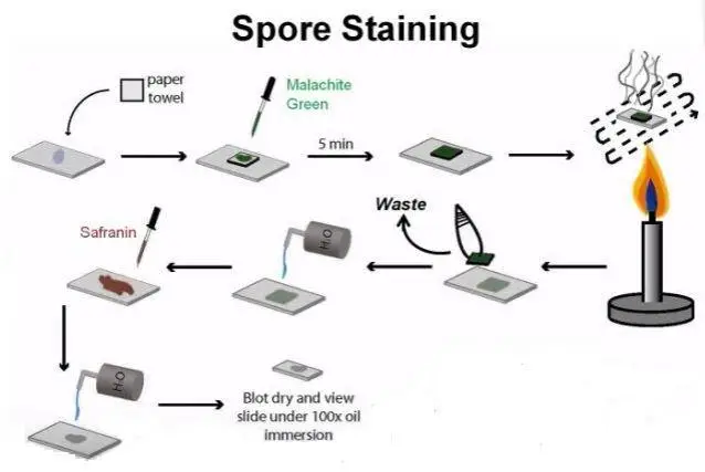
In the endospore staining lab, a sample of bacteria is first heat-fixed to a microscope slide. The heat-fixing process helps to preserve the sample and make it easier to work with. Next, the slide is flooded with a crystal violet solution, which stains the endospores a dark purple color. The slide is then washed with a decolorizing solution, which removes the crystal violet from the vegetative cells but leaves it behind in the endospores. Finally, the slide is flooded with a counterstain called safranin, which stains the vegetative cells a pink color.
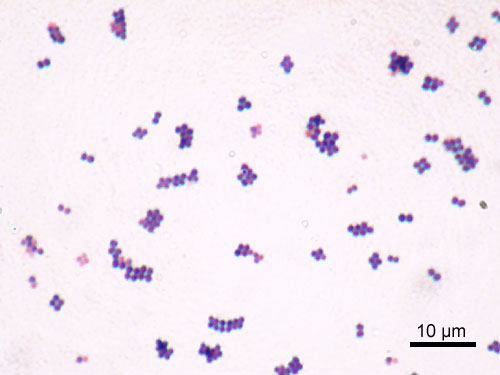
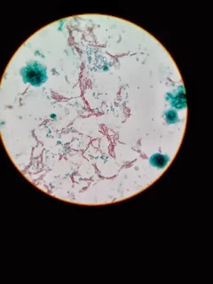
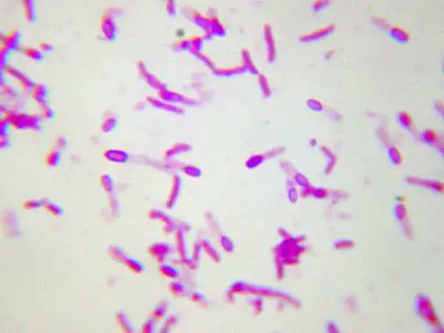
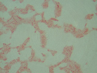
When viewed under a microscope, the endospores appear as dark purple structures within the pink-stained vegetative cells. This contrast makes it easy to identify and count the number of endospores present in the sample.
One important aspect of the endospore staining lab is the control sample. This is a separate slide that is treated in the same way as the experimental sample, but it does not contain any bacteria. The control sample serves as a reference point, allowing the researcher to confirm that the staining method is working properly.
The results of the endospore staining lab can be used in a number of different ways. For example, researchers can use the technique to identify the presence of endospores in food samples, to determine the effectiveness of disinfectants and antibiotics against endospores, and to study the survival mechanisms of endospores in different environments.
In conclusion, endospore staining is a valuable laboratory technique that allows researchers to visualize and study the highly resistant endospores produced by certain bacteria. This technique has a wide range of applications in the field of microbiology, including the identification and control of endospores in food and the development of new disinfectants and antibiotics.
ENDOSPORE STAIN
If the food is not properly processed, the endospores may germinate, produce toxins and cause food poisoning. . The Schaeffer- Fulton Spore staining technique is a differential staining method where malachite is the primary stain which stains the endospore green while safranin is the counter stain that stains the vegetative part of the bacterial pink such that both the structures can be viewed under the microscope with ease. The tests performed include: the Fermentation of Sugars Test sucrose, glucose, and lactose , the Urease Test, the Fermentation of Lactose Test, the Sulfide Indole Mobility SIM Test, the Nitrate Reduction Test, the Protein Hydrolysis Test, the Catalase Test, and the Cytochrome Oxidase Test. . Upon viewing my results it was determined that my colonies did not ferment mannitol.
Endospore Stain Lab blog.sigma-systems.com
Materials needed are Crystal Violet stain, Gram iodine stain, decolorizer, and the bacteria Staphylococcus epidermidis and Unknown Bacteria Essay 498 Words 2 Pages After incubation, a gram stain was performed one the colonies that were isolated. What are the functions of endospores in bacteria? Learn more For the endospore staining, Bacillus subtilis had a mixture of green and pink staining, indicating the presence of endospores. ENDOSPORE STAINING Endospores are stained bright green and vegetative cells are stained reddish thickness of the cell wall called peptidoglycan. Most capsules are composed of polysaccharides but a few are composed of polypeptides. Purpose The purpose of this lab is to visualize and study endospores prior to and after the counterstain. For broth cultures, no water is added. The sections that should be covered are outlined and a structure you could follow is proposed.
Complete Endospore Stain Lab

The endospore stain is used to determine if endospores are present in a bacterial smear. The smear will be flooded with malachite green 0. What is the stimulus for endospore production in bacteria? Acid-Fast Staining Two clean slides were obtained and labeled with the microorganism, date, and stain type. Due to this, Gram-negative cells can take on the red colored, Safranin stain. Iodine is then added which act as a mordant to trap the crystal violet dyes and form crystal violet-iodine complex. For instance, if you are working in Active Directory, you can create a new user account, set up a shared folder, or change to a different domain.