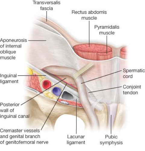
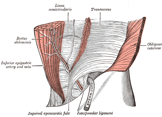
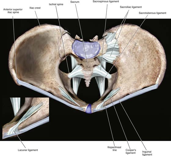
Lacunar ligament. Lacunar (Gimbernat's) ligament 2022-12-21

Lacunar ligament Rating: 6,6/10 1149 reviews

The lacunar ligament, also known as the ligamentum lacunare or ligamentum venosum, is a small, thin ligament located in the liver. It is an important anatomical structure that plays a crucial role in the functioning of the liver and its ability to filter blood and produce important enzymes and hormones.

The lacunar ligament is a long, narrow band of connective tissue that extends from the inferior vena cava (IVC) to the liver. It is located in the superior portion of the liver, just below the diaphragm. The IVC is a large vein that carries deoxygenated blood from the lower half of the body to the right atrium of the heart. The lacunar ligament forms a vital link between the IVC and the liver, allowing blood to flow freely between these two organs.

The lacunar ligament is an important part of the hepatic venous system, which is responsible for draining blood from the liver. It is composed of several smaller veins and tributaries that converge to form the lacunar ligament. These smaller veins and tributaries are called the hepatic veins, which carry oxygenated blood from the liver back to the heart.

The lacunar ligament is also closely associated with the portal vein, which is another important blood vessel in the liver. The portal vein carries blood from the intestines, spleen, and pancreas to the liver, where it is filtered and processed. The lacunar ligament helps to regulate the flow of blood between the portal vein and the hepatic veins, ensuring that the liver receives a steady supply of oxygenated blood while also preventing the build-up of excess pressure.

In addition to its role in the hepatic venous system, the lacunar ligament also plays a crucial role in the production of enzymes and hormones. The liver is responsible for producing a variety of enzymes and hormones that are essential for maintaining homeostasis in the body. These enzymes and hormones are produced by specialized cells called hepatocytes, which are found in the liver. The lacunar ligament helps to ensure that these hepatocytes receive an adequate supply of oxygen and nutrients, which are essential for their proper function.

Overall, the lacunar ligament is a small but essential structure that plays a vital role in the functioning of the liver. It helps to regulate the flow of blood between the IVC and the liver, as well as between the portal vein and the hepatic veins. It also helps to ensure that the hepatocytes receive an adequate supply of oxygen and nutrients, which are essential for the production of enzymes and hormones. Without the lacunar ligament, the liver would be unable to function properly, leading to serious health problems.
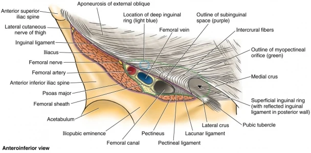
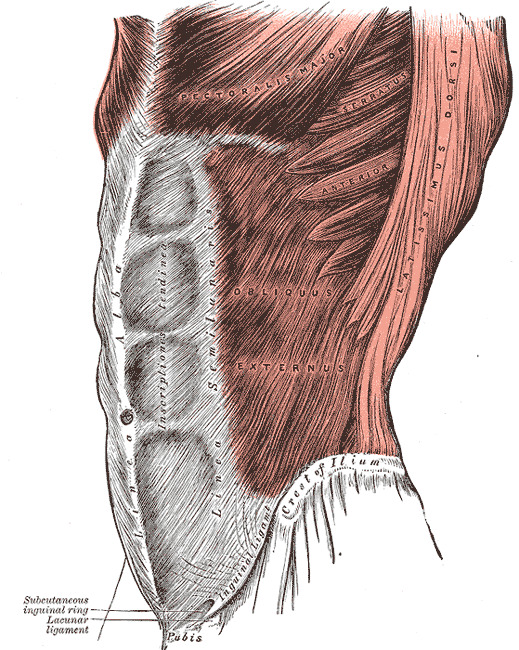
Inguinal ligament: Attachments, function and relations

lacunar ligament

The displacement of the tibia away from the femur is used to assess whether or not the ligament is torn. Carter never gone to India. It courses from the pre-auricular surface of the ilium to the third segment of the sacrum. Its composite bands extend between the adjoining fibular and tibial surfaces. Unlike the lateral epicondyle, the medial epicondyle has attachments to the joint capsule of the knee and by extension, to the medial meniscus.

Next

Lacunar Stroke: Symptoms, Treatments, and Long

lacunar ligament

Of note, the word crus pl. Lacunar stroke is a type of ischemic stroke that occurs when blood flow to one of the small arteries deep within the brain becomes blocked. The ligament extends from its basal attachment at the intertrochanteric line to its apex between the rim of the acetabulum and the anterior inferior iliac spine. We avoid using tertiary references. The information we provide is grounded on academic literature and peer-reviewed research. The ligament is made up of two bands that originate from the transverse processes of L5. While it does not integrate with the joint capsule, it blends with the tendon of biceps femoris to form its conjoint tendon.

Next

Lacunar (Gimbernat's) ligament

lacunar ligament

J Med Biogr 2000 8:206—212. It has attachments to the roughened posterior aspect of the patella, the distal apex of the patella as well as its margins. The stability of the pubic symphysis is reinforced by the superior and inferior arcuate pubic ligaments. Strokes that occur due to blockages in blood vessels within the brain are called ischemic strokes. For all his work and dedication, Dr.

Next

Lacunar ligament

lacunar ligament

Image property of: Artist: One of concers when using this technique is the presence of the. Inguinal hernias are classified as indirect or direct: Inguinal hernias that protrude through the deep inguinal ring and traverse the canal are known as indirect hernias. Tibial collateral ligament medial view Talocalcaneal ligaments Otherwise called the subtalar joint, the talocalcaneal joint represents the anteroposterior articulation between the talus and calcaneus. While direct hernias protrude through the superficial ring and emerge within the inguinal triangle of Hesselbach, inferomedial aspect of anterior abdominal wall. The tibiocalcaneal also known as the intermediate part takes a vertical route to insert along the upper border of sustentaculum tali a bony projection of the calcaneus. Click on the image for a larger depiction. Medical Terminology Daily MTD is a blog sponsored by Click on the link below to to the MTD newsletter.


Next

Ligaments of the lower limb: Pelvis, knee, ankle

lacunar ligament

The information we provide is grounded on academic literature and peer-reviewed research. The sacrotuberous ligament also serves as a point of attachment for the most caudal fibers of Sacrotuberous ligament dorsal view Obturator membrane Probably the most confined of all the ligaments of the pelvis is the obturator membrane. Philadelphia, PA: Churchill Livingstone: Elservier. If you think an article could be interesting to somebody else, feel free to forward the link of the article. They may also carry out a detailed neurological exam to see if there is any damage to the parts of the brain that control your bodily functions. It then travels across the sciatic notch to complete its connection to the falciform process. Additionally, it has cranial attachments to the calcaneofibular ligament of the lateral ligament of the ankle.

Next

Gimbernat’s ligament

lacunar ligament

In this maneuver, the patient is supine and the knee is flexed at 90 degrees. Note that the pre-auricular surface of the ilium is the area just anterior to the auricular surface, which is the area of articulation between the ilium and the sacrum. The ligament is the anterior cruciate, and the injurious force is often valgus in nature. Do not confuse this ligament with others with the same name found in the breast. Of note, the posterior ligament is a solitary band that travels superomedially from the posterior aspect of the fibular head to the lateral condyle of the tibia. Â Atlas Of Human Anatomy. Carter only received a one-time payment of 150 pounds.

Next

Pectineal ligament

lacunar ligament

Here is the final recap for this article. Essential Clinical Anatomy: Third Edition. Carter was relegated to the position of illustrator by Henry Gray and never saw the royalties that the book could have generated for him. If you have high blood pressure, heart disease, or diabetes, try to keep it under control and see your doctor regularly. Rehabilitation Lacunar stroke can result in some brain damage.

Next

Lacunar_ligament : definition of Lacunar_ligament and synonyms of Lacunar_ligament (English)

lacunar ligament

It courses obliquely in an inferomedial direction to insert onto the pubic tubercle of the pubic bone. Recall that fibrous bone to bone, similar to the way tendons attach muscles to bones. The pectineal ligament is one of the targets used to place staples or bone tacks to attach a mesh that reinforces the hernia repair. Carter never worked again with Gray, who died of smallpox only a few years later. It also takes a posteroinferior course originating just inferior to the adductor tubercle of the medial epicondyle of the femur to the medial epicondyle of the tibia. Patellar ligament Finally, the patellar ligament is a thick fibrous structure that connects the patella to the tibia.


Next

Pectineal (Cooper's) ligament

lacunar ligament

What Gimbernat did was to carefully cut the ligament from lateral to medial, enlarging the femoral ring and allowing reduction of the hernia. However, the anterior ligaments consist of about three bands that also take a superomedial course from the anterior part of the head of the fibula to the lateral condyle of the tibia. The ligament is triangular in nature and its apex attaches to the fovea of the head of the femur, while its base inserts around the acetabular notch at the center of the acetabulum. A stroke occurs when a blockage interrupts or prevents blood flow to the brain. The ligament then inserts into the tibial tuberosity on the anterior surface of the tibia. From here the pectineal ligament extends laterally as part of the This is important in the repair of inguinofemoral hernias though laparoscopic procedures.

Next

lacunar ligament

The anterior tibiotalar part terminates on the medial talar aspect of the bone that does not articulate with the navicular bone; while the posterior tibiotalar component travels posteromedially to inserts on the medial talar tubercle and the medial surface of the talus. The named ligaments involved in stabilizing this joint are the interosseous, medial, lateral and cervical ligaments. Passing through the inguinal canal are the Put that newly-acquired knowledge to the test with the quiz below! The injury can be clinically assessed using an anterior drawer test. It forms the posterior border of the femoral canal. Subsequently, pain and swelling will develop.

Next