Structure of fasciola hepatica. Fasciola Hepatica: Habitat, Structure and Life History 2022-12-20

Structure of fasciola hepatica Rating: 6,6/10 1137 reviews

Fasciola hepatica, also known as the liver fluke, is a parasitic flatworm that causes a disease known as fascioliasis in humans and animals. It is found in many parts of the world, including Europe, Africa, and parts of Asia and South America.

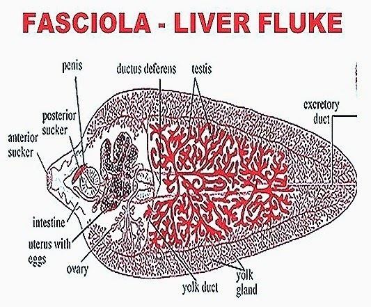
The structure of F. hepatica is complex, with several distinct regions that are important for its survival and reproduction. The head end of the fluke, known as the scolex, is characterized by a series of suckers and hooks that allow it to attach to the intestinal walls of its host. The body of the fluke is elongated and flattened, and is divided into three main regions: the neck, the ventral sucker, and the posterior end.

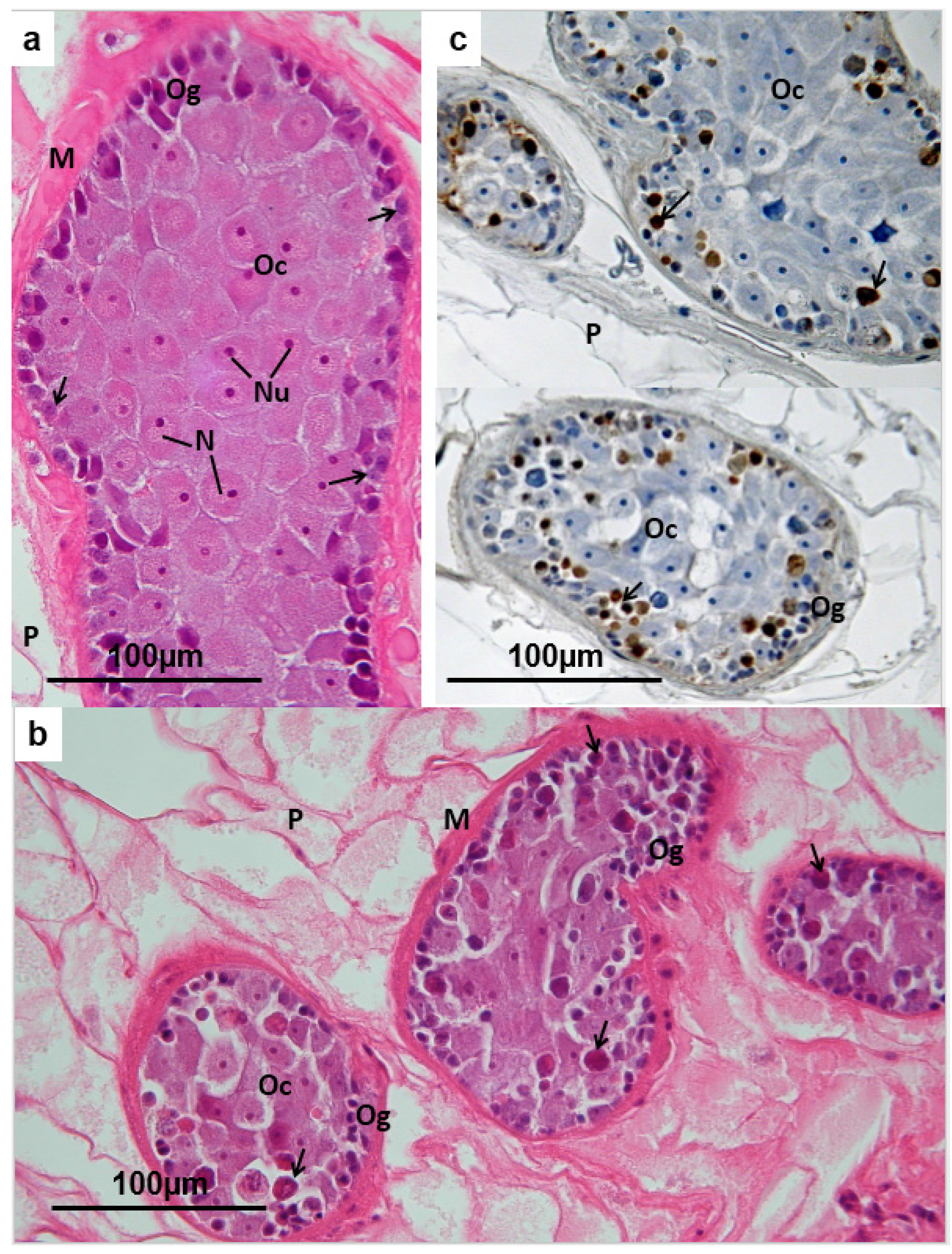
The neck region is located between the scolex and the ventral sucker, and is characterized by a series of muscles and reproductive organs. The ventral sucker is a circular region located near the midpoint of the body, and is used for attachment and movement. The posterior end of the fluke is characterized by the presence of a large digestive gland and a reproductive system, including both male and female organs.

F. hepatica is a hermaphroditic species, meaning that it has both male and female reproductive organs. The fluke is capable of self-fertilization, but it also has the ability to mate with other individuals. The eggs of the fluke are laid in the intestine of the host and are passed out in the feces. They hatch in water, and the larvae, known as miracidia, infect snails, which act as intermediate hosts. The larvae then develop into a form known as cercariae, which can infect mammals, including humans and livestock.

In conclusion, the structure of F. hepatica is complex, with several distinct regions that are important for its survival and reproduction. Understanding the anatomy and life cycle of this parasitic flatworm is important for the development of effective prevention and treatment strategies for fascioliasis.

Life Cycle of Fasciola Hepatica

structure of fasciola hepatica

The excretory system is protonephric type. It causes liver rot, liver cirrhosis, eosinophilia and anemia. The culture remained at 28 °C for a further 2 hours before cells were harvested by centrifugation 8000 × g, 20 minutes, 4 °C. Copulation, Fertilization and Capsule Formation: Copulation occurs in the bile duct of sheep. Once in the snail, the larvae reproduce and eventually release more larvae into the water. This method of development takes place in the formation of all larval stages during the life history. Expansion of the granular cushion, accompanied by exosmosis of salts and other materials from within the egg pushes off the operculum.

Next

Structure of Fasciola (With Diagram)

structure of fasciola hepatica

A birth pores in present lateral to and in proximity of the mouth. PROCHECK: a program to check the stereochemical quality of protein structures. The capsules which fall in water or damp places will develop at about 75°F. The first row consists of six plates 2 dorsal, 2 ventral and 2 lateral , the second row of 6 cells 3 dorsal and 3 ventral , the third row or 3 cells 1 dorsal, 2 ventrolateral , the fourth row of 4 cells 2 right and 2 left and the fifth row of 2 cells 1 right and 1 left only. Human Fascioliasis: A re-emerging disease in upper Egypt. Visual automated macromolecular model building.

Next

RCSB PDB

structure of fasciola hepatica

It serves the purpose of storing sperms. There are further non-bonded contributions to the binding from the amino acid residues Phe11, Phe13, Gly15, Pro54 and Tyr110. Towards the posterior side are some propagatory cells germ cells , some of which may have divided to form germ balls which are developing embryos. In fact, the mesenchyme forms a packing material between the muscle layer and internal organs. It reaches the liver through the hepatic portal system and starts its existence inside the bile passage.

Next

X

structure of fasciola hepatica

Sporocyst Larva: Miracidium loses its apical gland, penetration grand, brain and eyespots and changes into a sac­like sporocyst larva. The bodywall of redia is comprised of a thick cuticle, a squamous epithelium, a muscular and a mesenchyma layer. A pair of lateral cerebral and a ventral ganglia are present over the cerebral ring. Anteriorly, the two vasa deferentia join to form a median vesicula seminis, from which nans an ejaculatory duct, opening at the tip of the cirrus or penis, which, in turn, opens in the genital atrium. The terminal part of uterus has muscular walls, referred to as metraterm which ejects the eggs and also sometimes receives the cirrus during copulation. In the CGL complex the Tyr10 OH is 2.

Next

Fasciola: Digestive System and Excretory System

structure of fasciola hepatica

The 2Fo - Fc map blue is contoured at 1. ADVERTISEMENTS: At 30°C the encapsulated embryo differentiates into miracidium larva within 8 days. Metacercariae excsts in the duodenum or jejunum and liberate the juvenile fluke. Digestive System of Fasciola Hepatica : i Alimentary Canal: The oral sucker encloses a ventral mouth which leads into a funnel- shaped mouth cavity, followed by a round muscular pharynx with thick walls, and a small lumen. Just behind the collar is a birth pore through which next generation of larvae come out.

Next

Structure of Liver Fluke (Fasciola Hepatica)

structure of fasciola hepatica

These open on the surface by a common pore. In addition, there is a pair of large pigmented eyespots, a large larval brain and a simple nervous system in the anterior part of body. Ingestion of free metacercariae floating on water may also be a possible mode of transmission. These glands open into the ejaculatory duct and their secretion alkaline helps in free movement of sperms during copulation. During summer months when the food is sufficiently available, mother radiae mature in 12-18 days. Retrieved 11 January 2022.


Next

Fasciola Hepatica: Habitat, Structure and Life History

structure of fasciola hepatica

There are two muscular suckers an oral sucker at the anterior end encircling the mouth, and a large ventral sucker or acetabulum situated mid-ventrally about 3 to 4 mm behind the oral sucker. Miracidium Larva of Fasciola 7. Its secondary hosts are either Planorbis sps, Bulinus sps. In the GSSG complex subunits the Tyr10 OH atom is closer to the Sγ atoms of the GSSG complex than the Sγ atom of the GSH. It is a free swimming larval stage.

Next

Fasciola hepatica

structure of fasciola hepatica

Later the propagatory cell divides into two cells, one of which forms the endoderm and mesoderm of the larva, and the other forms a mass of germ cells at the posterior end of the larva. Nervous System of Fasciola Hepatica : A nerve ring surrounds the oesophagus, it has a pair of cerebral ganglia dorsolaterally, and a ventral ganglion below the oesophagus. Epidemiology They are worldwide distribution in distribution and economic importance in livestock raising. A system of excretory vessel is present. Full size image Quaternary Structure The FhGST-S1 forms dimers related by a molecular dyad with approximate dimensions 46 Å × 53 Å × 50 Å. Now the larva is known as metacercaria.

Next

Fasciola: Introduction, Morphology, Life Cycle, Pathogenecity, Laboratory

structure of fasciola hepatica

Molecular replacement with MOLREP. Nerve cords are connected by transverse commissures and they give out many small branches, some of which form plexuses. These open on the apical lobe or cephalic cone. Cilia are absent in adult flukes. Bioinformatics 24, 2780—2781 2008. Sperms travel down the uterus. The internal cavity contains germ cells.

Next