The posterior horns of the spinal cord contain. Posterior horn of spinal cord 2022-12-15
The posterior horns of the spinal cord contain Rating:
8,2/10
1450
reviews
The posterior horns of the spinal cord are a vital component of the central nervous system. Located in the posterior (back) portion of the spinal cord, these structures are responsible for receiving sensory input from the body and transmitting it to the brain for processing.
The posterior horns contain a collection of cells called interneurons, which are responsible for transmitting sensory information from the body to the brain. This information includes things like touch, temperature, pain, and proprioception (the sense of body position and movement).
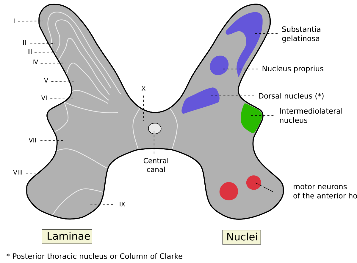
The cells within the posterior horns are organized into layers called laminae, with each layer responsible for processing a specific type of sensory information. For example, the laminae responsible for processing pain are called the substantia gelatinosa. These cells receive input from sensory neurons located in the body's tissues and transmit this information to the brain via the posterior horn.
In addition to interneurons, the posterior horns also contain a type of neuron called a motor neuron, which is responsible for transmitting signals from the brain to the body's muscles and organs. These signals control movements such as walking, running, and even breathing.
Overall, the posterior horns of the spinal cord play a crucial role in the body's ability to sense and respond to its environment. Without them, we would be unable to feel pain, touch, or sense our body's movements, which would greatly impair our ability to function in daily life.
Dee wants the old quilts for several reasons but mainly because she wants to display them as part of her? Cervical - thoracic - lumbar - sacral - coccygeal C. Two arms located at the front of the spinal cord, central grey matter are called ventral horns. She is much more closely tied to her family, and is making plans to marry. This article covers the key anatomy of the spinal cord and its functions. Kumar MD, in Handbook on Opium, 2022 13. What makes the quilts valuable to Dee? What is posterior cord syndrome? Diffusion depends on molecular weight, concentration gradient, ionic-to-nonionic ratio, and, most important, lipid solubility.
What do the posterior horns of the spinal cord contain? – Find what come to your mind
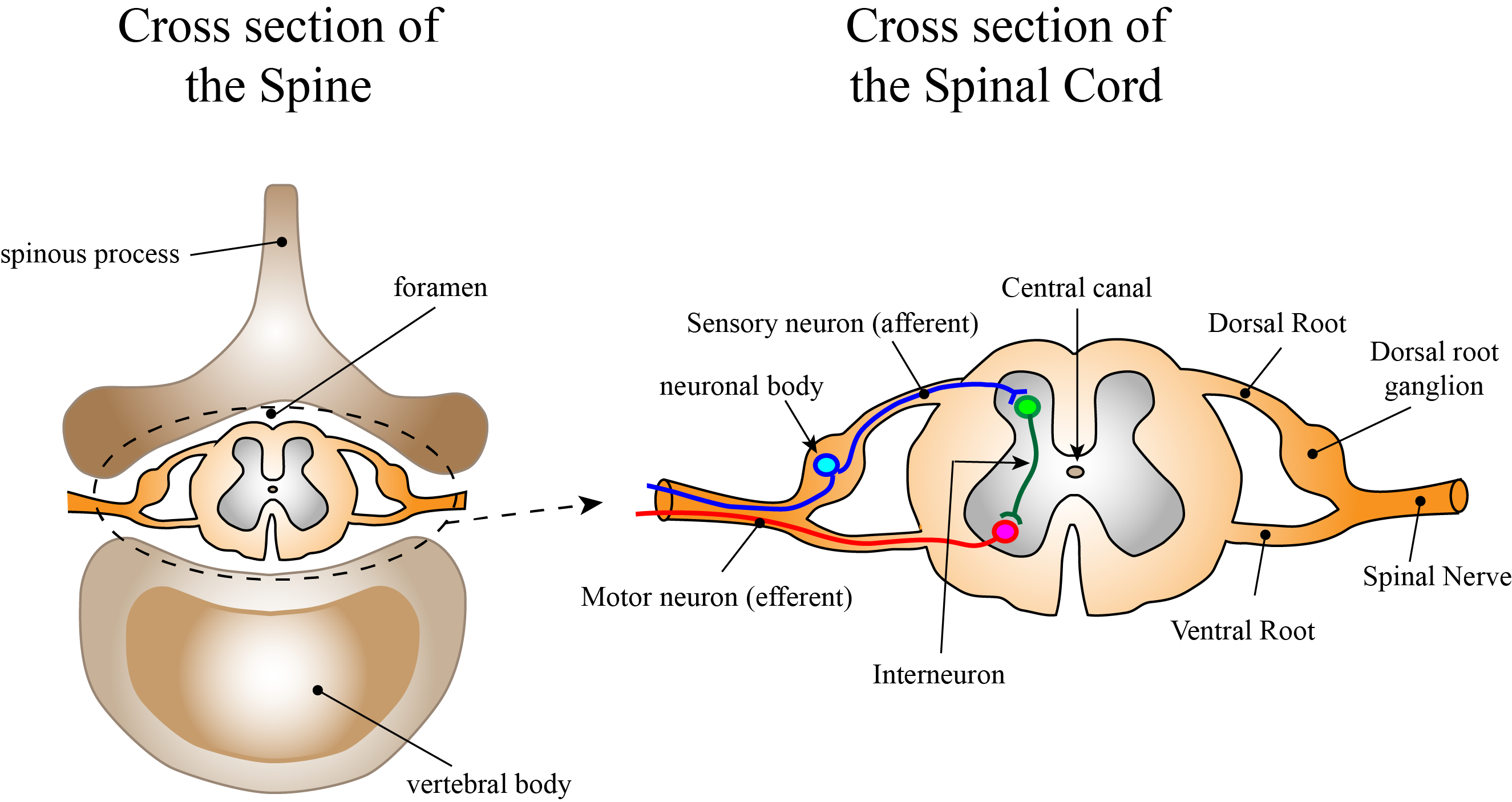
The motor neuron that causes contraction of the big toe, for example, is located in the sacral spinal cord. Whereas the brain develops out of expansions of the neural tube into primary and then secondary vesicles, the spinal cord maintains the tube structure and is only specialized into certain regions. C the spinal cord would not be able to process information at that level. Cookies allow us to analyze and store information such as the characteristics of your device as well as certain personal data e. It receives several types of sensory information from the body, including light touch, proprioception, and vibration. Which of the following is true regarding an epidural block? What specifically is found in the anterior gray horn of the spinal cord? What happens if you sever your spinal cord? Why does Dee want the old quilts in everyday use? The dorsal root ganglia are collections of sensory cell bodies with axons extending peripherally as well as a central process which passes into the spinal cord in the region of the posterior horn of grey matter and makes appropriate central connections.
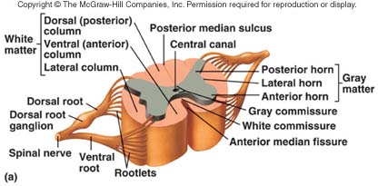
The spinal cord is a cylindrical structure of nervous tissue composed of white and gray matter, is uniformly organized and is divided into four regions: cervical C , thoracic T , lumbar L and sacral S , Figure 3. On the whole, the posterior regions are responsible for sensory functions and the anterior regions are associated with motor functions. Between the two posterior horns of gray matter are the posterior columns. In a pig model using a microdialysis technique, which enabled continuous sampling of opioid concentrations in the epidural and intrathecal spaces, Bernards et al 55 showed that there is a strong correlation between lipid solubility of a drug and 1 the time spent in the epidural space and 2 the terminal elimination half-life i. What causes anterior horn cell disease? Somatosensory imprinting depends on the tactile input that is associated with spontaneous movements that occur during sleep and results in elimination of inappropriate connections and establishment of appropriate connections.
C Tina has a spinal injury in the lumbar region. The reflex action you relied upon is most correctly known as a n A. Kenhub does not provide medical advice. The grey matter also extends from the brain into the spinal cord. Can you walk again after spinal cord injury? Rapamycin could effectively block mechanical and thermal hyperalgesia in CFA-, carrageenan-, and formalin-induced inflammatory pain Liang et al. Thus, opioids play a key role in the modulation and control of pain responses.
Schouenborg, in The Senses: A Comprehensive Reference, 2008 The organization of the somatosensory input to the dorsal horn of the spinal cord has for a long time been viewed as a somatotopical map of the body that is adult-like already at birth. Ketamine can be of particular benefit in clinical situations where the pain may be of a mixed nociceptive and neuropathic nature e. Read also How often do you trim the roots of a bonsai? The term posterior horn also dorsal horn, posterior cornu, dorsal cornu may refer to: … Posterior horn of spinal cord, the dorsal towards the back grey matter section of the spinal cord that receives several types of sensory information from the body including light touch, proprioception, and vibration. However, encephalopathy is becoming an increasingly recognized complication of diabetes, largely within the context of the cognitive impairments seen in both humans Biessels and Kappelle, 2005; Biessels et al. The Everyday Use quotes below all refer to the symbol of Quilts. E all of the above If the dorsal root of a spinal nerve is severed, A output to skeletal muscles would be blocked. The hydrophilic opioids, such as morphine, are slowly removed from the CSF, leading to rostral spread of relatively high concentrations of the drug.
They receive collateral or direct terminations of dorsal root fibers. Where are the dorsal horns? It receives several types of sensory information from the body, including fine touch, proprioception, and vibration. The spinal cord is not the full length of the vertebral column because the spinal cord does not grow significantly longer after the first or second year, but the skeleton continues to grow. Nociceptors directly synapse with inhibitory interneurons that are characterized by the production of -aminobutyric acid GABA Ch. Read also What is the demand curve for a perfectly competitive firm? DAMIAN MURPHY, in Postoperative Pain Management, 2006 SPINAL ROUTE: EPIDURAL AND INTRATHECAL Opioid receptors located in the dorsal horn of the spinal cord are not confined to the superficial layers but are also present in the substantia gelatinosa, implying that spinally administered opioids must diffuse into the deep layers of the spinal cord in order to induce analgesia. For the two older women, heritage means passing down skills? Our recent work demonstrated that intrathecal rapamycin not only attenuated neuropathic pain but also delayed initiation of morphine tolerance and hyperalgesia in neuropathic pain rats Xu et al.
A spinal cord injury is a life changing and devastating event. These results suggest that A Tina has injured one of her descending nerve tracts. Dextromethorphan, a component of cough mixtures, is an alternative, although less potent, NMDA receptor antagonist that has been shown to reduce opioid requirements following oral or abdominal surgery. The anterior horn sends out motor signals to the skeletal muscles. Read also Should you cover outdoor water faucets? Review article the dorsal horn of the spinal cord. Acute flaccid paralysis secondary to anterior horn cell disease may be associated with other enteroviruses e.
As a result, which of the following would you expect? Where do you find quilts in everyday life? Does a spinal cord injury shorten your life? Inhibition of the mTORC1 pathway relieved morphine tolerance and hyperalgesia. The exact site of action of lipophilic agents administered epidurally is uncertain, because plasma levels of these drugs, analgesia, and side effects are similar for both epidural and intravenous administrations. Are there cell bodies in the dorsal horn? The ventral horns contains the cell bodies of motor neurons that send axons via the ventral roots of the spinal nerves to terminate on striated muscles. This comes from the initial development of the spinal cord, which is divided into the basal plate and the alar plate. The lateral horn, which is only found in the thoracic, upper lumbar, and sacral regions, is the central component of the sympathetic division of the autonomic nervous system. Read also Can products be copyrighted? To a great extent, the quilt embodies the personalized connection that both mother and daughter share to one another and their past.