Tibia osteology. Tibia: Anatomy and clinical notes 2022-12-15

Tibia osteology Rating: 6,5/10 1184 reviews

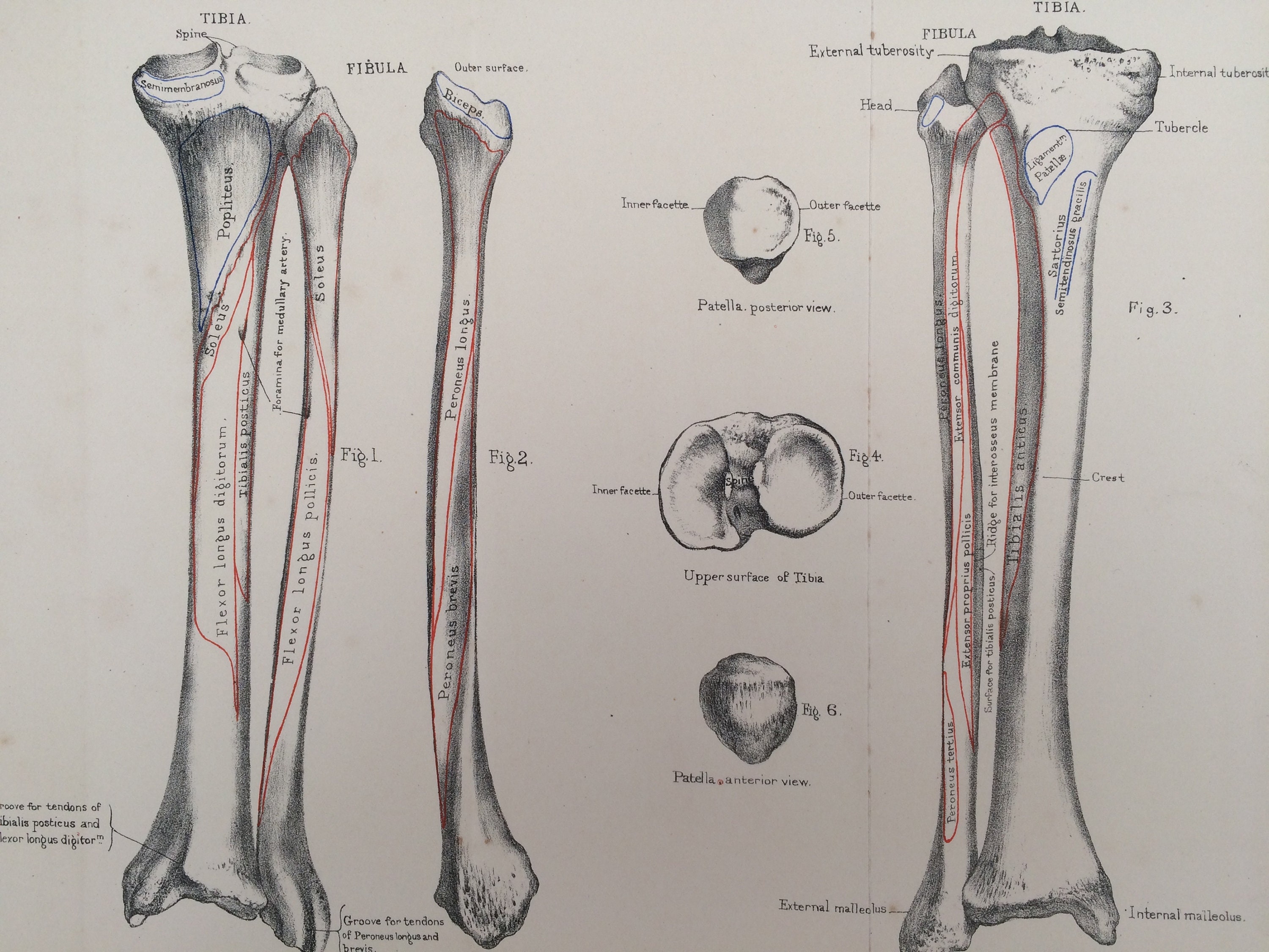
The tibia, also known as the shin bone, is a long bone located in the lower leg between the knee and the ankle. It is the larger of the two bones in the leg, with the other being the fibula. The tibia serves several important functions in the body, including bearing weight, providing stability, and allowing for movement of the leg.

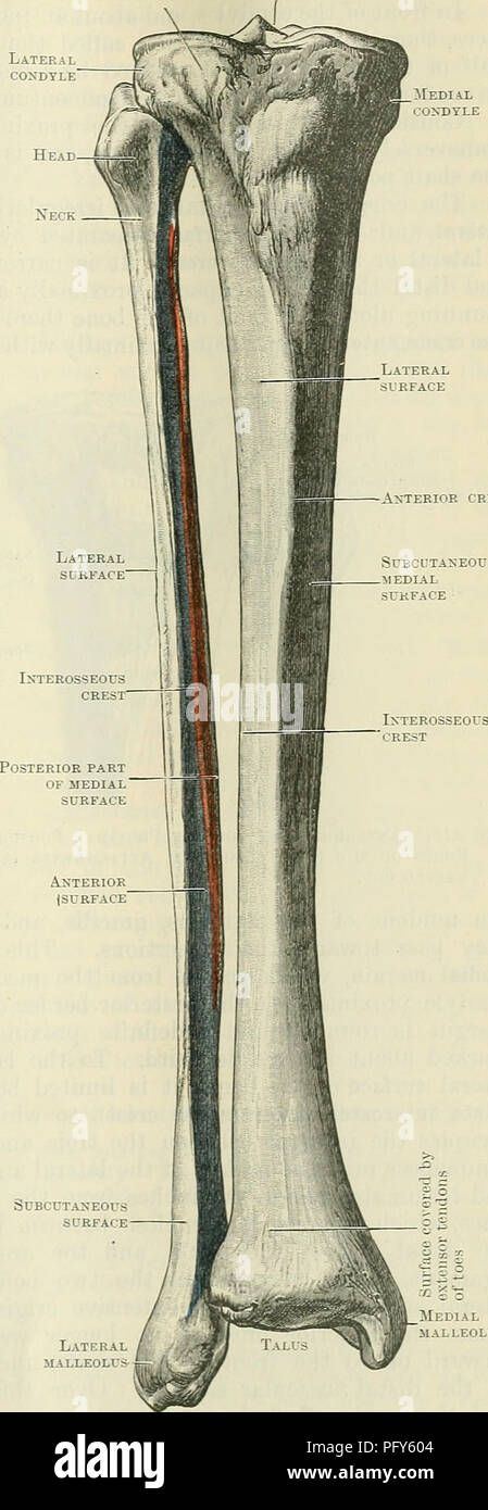
Osteology is the study of the bones of the body, and the tibia is an important bone to consider when studying the osteology of the leg. The tibia is a long bone with a cylindrical shape and a pointed end at the top, known as the proximal end. The bottom end of the tibia, known as the distal end, is wider and flattened. The tibia is covered in a thin layer of articular cartilage, which helps to reduce friction and wear on the bone during movement.

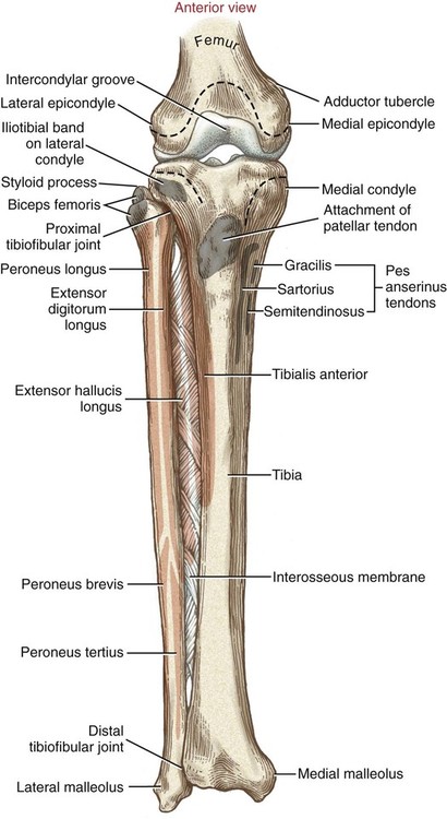
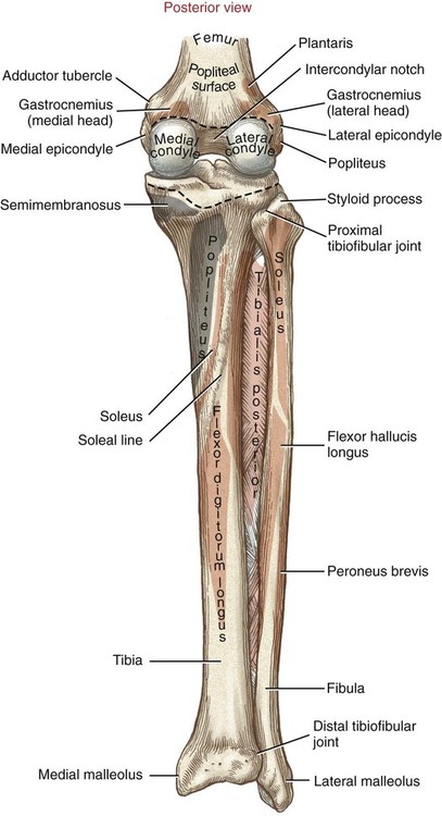
The tibia has several bony protuberances, or prominences, that serve various functions. The medial malleolus is a bony prominence on the inner side of the ankle that forms the bony prominence of the ankle. The lateral malleolus is a similar bony prominence on the outer side of the ankle. These prominences provide stability and support for the ankle joint.

The tibia also has several important articulations, or joints, with other bones in the body. The proximal end of the tibia articulates with the femur, the bone of the upper leg, to form the knee joint. The distal end of the tibia articulates with the talus bone of the foot to form the ankle joint. These joints allow for movement of the leg, including flexion and extension of the knee and plantar and dorsiflexion of the ankle.

In addition to its role in movement and stability, the tibia also plays a role in the circulation of blood in the body. The tibia contains several blood vessels and nerves that supply blood and sensation to the lower leg and foot. The tibia is also home to the tibialis anterior muscle, which is responsible for moving the foot upward and outward.

Overall, the tibia is an important bone that plays a vital role in the movement and stability of the leg, as well as in the circulation of blood and sensation in the lower leg and foot. Understanding the osteology of the tibia is important for understanding the overall anatomy and function of the leg.

Tibia: Anatomy and clinical notes

tibia osteology

The fibula does not articulate with the femur, so it bears very little weight. The posterior margin runs the length of the dorsal shaft surface. The medial malleolus projects distally from the medial aspect of the distal tibia. The proximal fibular articular facet is perched just below the lateral condyle on the dorsal aspect of the proximal tibia. Fractures are easily seen on an X-ray of the affected leg.

Next

Osteology, Arthrology and Biomechanics: Tibia

tibia osteology

From this view it can be seen that the apex is nonarticular. The dorsal surface is a large articular surface with a medial articular facet and a lateral articular facet for the patellar surface of the femur. The anterior intercondylar area houses the attachment of the The diaphysis of the tibia is triangular in cross section and has medial, lateral and posterior surfaces, separated by the anterior, lateral interosseous and medial borders. The fibular notch on the lateral side of the distal fibula is the distal articular surface for the fibula. Two depressions for the attachment of the cruciate ligaments of the knee are located just anterior and posterior to these tubercules. Its most vulnerable part is the tibial shaft and it generally takes a strong force to break this bone. This lobe is on the ventral side.

Next

Osteology: The Tibia

tibia osteology

It is the largest bone in the human body. The posterior surface is bound by the interosseous and medial margins and features the soleal line crossing this surface diagonally. The articular surface of the femoral head is disrupted by the fovea capitis, an excavated depression in the otherwise smooth surface. Test yourself and learn them very fast using active recall and Kenhub's Distal part At the distal end of the tibia, the bone has changed shaped from triangular in cross section to rectangular in cross section. Instead, the lateral margin extends to the medial intercondylar tubercle. Fibular shaft fragments can be quite difficult to side without comparative material.

Next

Tibia

tibia osteology

The shaft tapers distally, then expands slightly to form the articular surface for the talus at the ankle joint. The medially located interosseous margin is more rounded than the anterior margin. Â Note that the lateral and medial menisci are the pads of Synonyms: Eminentia intercondylaris The superior surfaces of the condyles are flattened and together they form the superior articular surface called the tibial plateau. The interosseous margin is lateral, and is offset dorsally. The medial margin extends to the lateral intercondylar tubercle. The proximal femur is dominated by its massive, nearly spherical head.

Next

Osteology: Leg

tibia osteology

Thus, because males and females differ in average body mass a condition known as sexual dimorphism , the diameter of the femoral head alone is a fairly good predictor of sex. It articulates with the talus within the ankle joint. Two roughed trochanters on the proximal femur provide important muscle attachment sites. The leg, or lower limb, is comprised of three major bones: the femur, tibia, and fibula. Clinically Oriented Anatomy 7th ed. It articulates proximally and distally with the tibia and forms the lateral bony prominence that is palpable at the ankle, where it articulates with the talus. This video covers the osteology of the tibia.


Next

High Tibial Osteotomy Knee Surgery and Realignment

tibia osteology

Synonyms: Tibial bone The tibia shin bone is a long bone of the Like other long bones, there are three parts of the tibia: proximal, shaft, and distal. The gluteus medius and gluteus minimus muscles take origin here and serve as critical hip stabilizers during walking. The apices of this triangle correspond with the medial margin, interosseous margin, and the anterior margin of the tibia. Its articular surfaces are the superior surfaces of lateral and medial condyles of the tibia, and the inferior surfaces of the lateral and medial condyles of the femur. An osteotomy may also be performed in conjunction with other joint preservation procedures in order to allow for cartilage repair tissue to grow without being subjected to excessive pressure. Stewart 1979 used 50 male and 50 female skeletons of known sex to show that 93% of skeletons in his sample could be correctly assigned to sex using femoral head diameter alone see Figure.

Next

tibia osteology

The lateral articular facet is larger than the medial facet. On the other hand, the superior surface of the lateral condyle is pretty much a mirror image of the medial condyle. The proximal ossification center appears at birth and fuses in the sixteenth year in females and the eighteenth year in males. Viewed from the ventral aspect, the patella is shaped like a clam, with a distally pointed apex and a rounded base. The patella is the largest sesamoid bone in the human body. Just posterior to this surface is the maleolar fossa, which serves at the attachment point for several ankle ligaments.

Next

tibia osteology

Of the three borders, the anterior border is the most prominent. This procedure is typically reserved for younger patients with pain resulting from instability and malalignment. The malleoli are bony prominences on the outer marigins of the sagittal grooves. The greater trochanter points superiorly and the lesser trochanter is dorsal. These tubercles form the intercondylar eminence, which is bordered by the anterior and posterior intercondylar areas.

Next

tibia osteology

The proximal fibula has a bulbous head with a superiorly projecting apex on the lateral side. The two trochanters are united by an intertrochanteric crest on the dorsal surface of the proximal femur and an intertrochanteric line on the ventral surface. These muscles are ankle dorsiflexors. In addition, there is also a portion showing major motions and joint involvements as well as some clinical correlations affecting the tibia. For some patients who have knee arthritis, this surgery can delay or prevent the need for a Osteoarthritis and Malalignment of the Knee With each step you take, forces equal to 3 to 8 times your body weight travel between the femur thighbone and tibia shin bone in your knee. It is a sheet of fibrous tissue that joins the tibia and fibula in the tibiofibular syndesmosis. The sharp ventral margin of the tibial shaft is easily palpated in the living person; this is the familiar shin bone.

Next

tibia osteology

The soleal line slants from lateral to medial as it plunges distally. At the distal end of the tibia is another major joint: the ankle joint. It is round in shape, somewhat convex, and articulates with the lateral condyle of the femur. For shaft fragments, the linea aspera is dorsal, and the shaft widens medially-laterally on the dorsal end. The joint where the femur articulates with the tibia is the knee.

Next