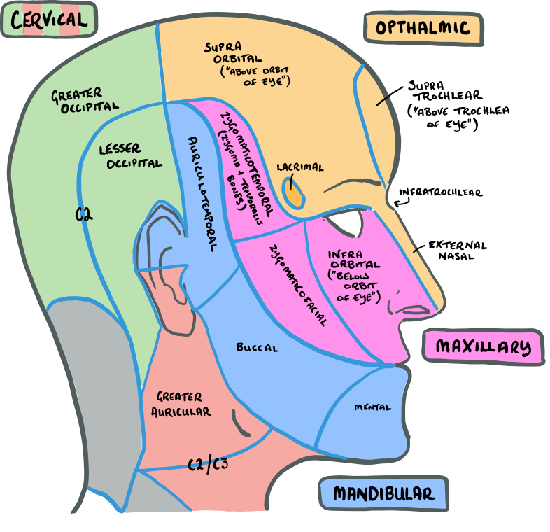
The trigeminal nerve is the fifth cranial nerve, also known as the mandibular nerve or the fifth cranial nerve. It is the largest of the cranial nerves and is responsible for transmitting sensory information from the face and head to the brain. The trigeminal nerve is made up of three branches: the ophthalmic branch, the maxillary branch, and the mandibular branch.
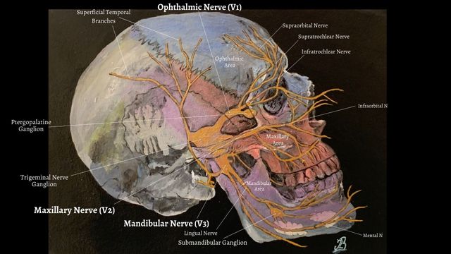
The ophthalmic branch, also known as the V1 branch, originates from the trigeminal ganglion and travels through the orbital fissure to reach the orbit of the eye. It innervates the skin of the forehead and scalp, as well as the conjunctiva and mucous membranes of the eye.
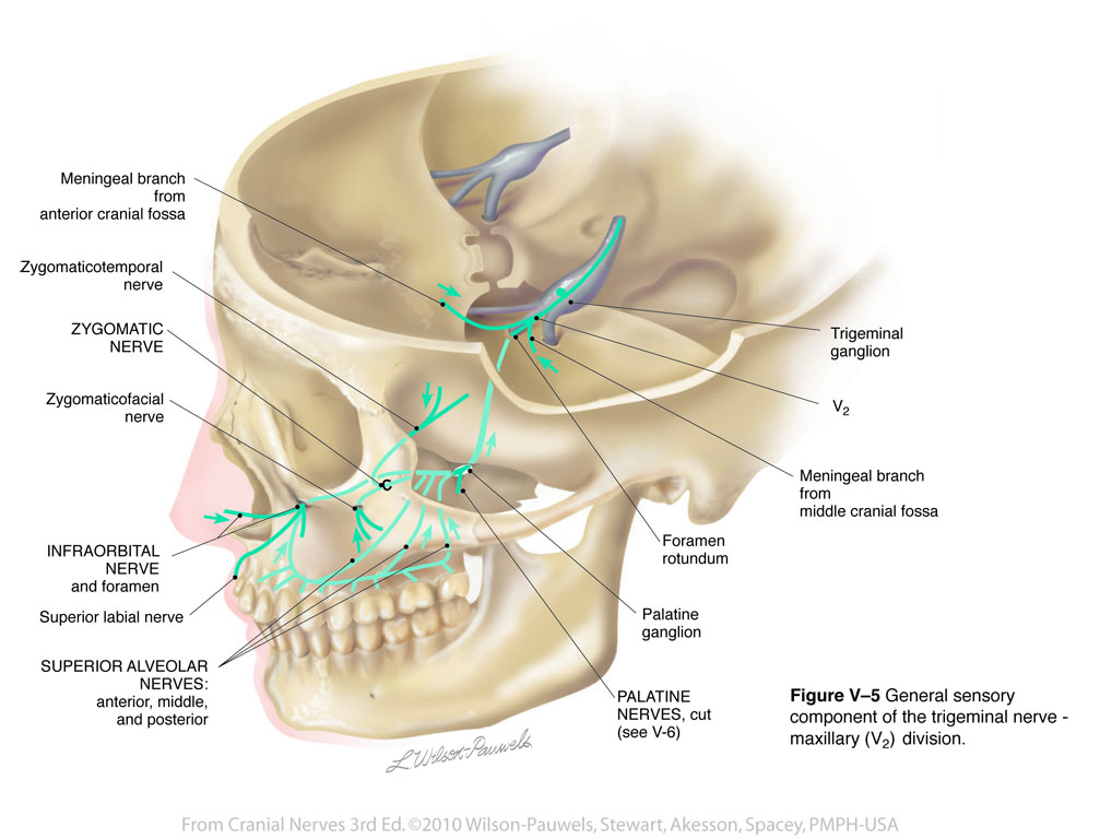
The maxillary branch, or V2 branch, also originates from the trigeminal ganglion and travels through the foramen rotundum to reach the pterygopalatine fossa. It innervates the skin of the cheek, upper lip, and nose, as well as the mucous membranes of the sinuses and the palate.
The mandibular branch, or V3 branch, is the largest of the three branches and originates from the trigeminal ganglion and travels through the foramen ovale to reach the mandible. It innervates the skin of the lower lip, chin, and lower jaw, as well as the teeth and gums.
The trigeminal nerve is involved in a number of important functions, including the sense of touch, pain, and temperature sensation in the face and head. It also plays a role in the reflexes involved in biting and chewing.
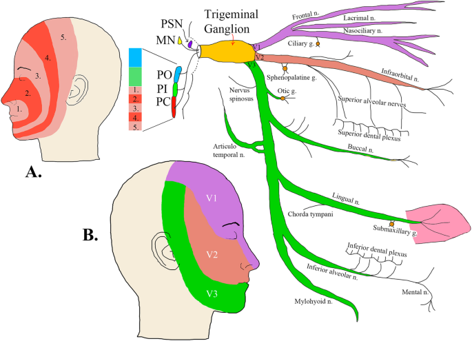
The trigeminal nerve pathway begins in the trigeminal ganglion, a group of sensory neurons located in the pons of the brainstem. From there, the nerve branches out to the three branches mentioned above, which innervate specific areas of the face and head. The sensory information transmitted by the trigeminal nerve is then relayed to the brain through the thalamus, where it is processed and interpreted.
In summary, the trigeminal nerve is a crucial cranial nerve that plays a vital role in the sensory functions of the face and head. Its pathway begins in the trigeminal ganglion and branches out to the ophthalmic, maxillary, and mandibular branches, which innervate specific areas of the face and head. The sensory information transmitted by the trigeminal nerve is then relayed to the brain for interpretation.
The Trigeminal Nerve (CN V)
Maxillary refers to the upper jaw. A major branch of this plexus, the mental nerve, supplies the skin and mucous membranes of the lower lip, skin of the chin, and the gingiva of the lower teeth. A study of 63 cases. Nerves that send sensory information have sensory functions. Waldman, in Pain Management Second Edition , 2011 Role of the Trigeminal System The trigeminal nerve is the largest and the most complex of the cranial nerves, containing sensory and motor fibers.
Some people need a nerve graft to replace the damaged nerve with a healthy one. Dental procedures and other injuries can cause numbness, or trigeminal neuropathy. Recall that prior to every clinical examination; the clinician should obtain informed consent from the patient. On the other hand, the peripheral branches originate from the neuromuscular spindle apparatus within the muscles of mastication, as well as from other proprioceptive points in the CN V3 , while those arising from the upper jaw gain access to the nucleus via the maxillary division of the trigeminal nerve CN V2. In addition, the tensor tympani, tensor veli palatini, anterior belly of the digastric, and mylohyoid muscle are also derived from first arch mesoderm. In the corneal reflex, the ophthalmic nerve acts as the afferent limb - detecting the stimuli.
Nevertheless, the examination requires inspection of the muscles. If one side is weak, the intact pterygoid muscles will push the weak muscles, resulting in a deviation toward the weak side. Sensory: The three terminal branches of CN V innervate the skin, mucous membranes and sinuses of the face. Once formed, the ophthalmic nerve also receives its meningeal tributary from the dura of the anterior cranial fossa. Functional MRI imaging of a defined stimulus for example, stroking the skin with a toothbrush "lights up" a single focus in SI and two foci in SII.
Trigeminal nerve (CN V): Anatomy, function and branches
Proceed to examine each trigeminal division by lightly tapping the cotton wisp on the territory supplied by each branch of the trigeminal nerve. Clinical examination Since the trigeminal nerve supplies motor and carries sensory stimuli, clinical examination of this nerve should evaluate the integrity of these modalities with regards to these stimuli. The peripheral aspect of the trigeminal ganglion gives rise to 3 divisions: ophthalmic V1 , maxillary V2 and mandibular V3. But note that CN V is NOT part of the cranial outflow of PNS supply Anatomical Course The trigeminal nerve originates from three sensory nuclei mesencephalic, principal sensory, spinal nuclei of trigeminal nerve and one motor nucleus motor nucleus of the trigeminal nerve extending from the midbrain to the medulla. Avoid applying too much pressure as the patient may misinterpret the pressure sensation for the light touch. Continuous pain, sometimes of a burning quality, is characteristic of posttraumatic neuropathy and is a common feature in postherpetic neuralgia. It tends to occur more often over time.
The Trigeminal Nerve: Anatomy, Function, and Treatment
 Motor examination Considering that temporalis and masseter are the only two superficial muscles of mastication, the test is usually limited. The lancinating, excruciating pain of trigeminal neuralgia must be distinguished from the more tolerable yet disturbing paresthesias of trigeminal sensory neuropathy. In the middle cranial fossa, the sensory root expands into the trigeminal ganglion. Divisions and merging of nerve branches can occur more distally closer to the skin or more proximally closer to the nerve root in the brain than expected. Pain can also be achy or burning.
In other cases, injecting a numbing agent into the nerve can help. The maxillary nerves extend to your cheeks, nose, lower eyelids and upper lip and gums. The central processing of pain-temperature information differs from the processing of touch-position information. Another part of the sensory test that is not frequently performed is the nasal tickle test. Their distribution pattern is similar to the dermatome supply of spinal nerves except there is little overlap in the supply of the divisions.
The fibers access the ventral posteromedial nucleus of the thalamus, after which third order neurons ascend through the internal capsule to gain access to Brodmann area 3, 1, 2 i. TN can cause long-term difficulties with chewing and speaking. The motor nucleus continues to form a motor root. Within the mandibular canal, the inferior alveolar nerve forms the inferior dental plexus, which innervates the lower teeth. Touch-position input comes to attention immediately, but pain-temperature input reaches the level of consciousness after a delay; when a person steps on a pin, the awareness of stepping on something is immediate but the pain associated with it is delayed.
The trigeminal nerve can also be a source of intense pain for some people. Based on symptoms, NOP may be divided into two broad categories: paroxysmal and continuous. These include the muscles of mastication , as well as the mylohyoid, anterior belly of the The motor nucleus receives extensive bilateral corticobulbar from the cerebral cortices to the cranial nerve nucleus , and rubrobulbar from the main sensory and mesencephalic nuclei that equally contribute to the regulation of the motor nucleus. These muscles are located in the jaw and their coordinated movement controls chewing. The muscles formed by mesodermal elements of the first branchial arch include the muscles of mastication: the temporalis, masseter, and medial and lateral pterygoid muscles. Then, touch the patient with the cotton wisp on an exposed area of skin, in the same manner, you would during the evaluation.
Check out our Ophthalmic division CN V1 The sensory nerve that carries afferent stimuli of pain, light touch, and temperature from the upper eyelids and supraorbital region of the face, up to the vertex of the head. The information we provide is grounded on academic literature and peer-reviewed research. Many things can trigger the pain, including touching the face, shaving, eating, yawning, or talking. It exits the middle cranial fossa via the foramen rotundum and crosses the pterygopalatine fossa where it is amenable to blockade by trigeminal nerve block via the coronoid approach. Head Trauma A traumatic injury can cause damage to the trigeminal nerve.
Atlas Of Human Anatomy. All 12 cranial nerves 12 in each side emerge from the brainstem. The present general understanding is that pain-temperature information from all areas of the human body is represented in the spinal cord and brainstem in an ascending, Within the spinal trigeminal nucleus, information is represented in a layered, or "onion-skin" fashion. The fibres supply parasympathetic innervation to the lacrimal gland. The brainstem is the lower part of the brain that serves as the physical connection between the spinal cord and the cerebral cortex of the brain. Descriptive terms—tingling, pins-and-needles sensation, burning, boring, and stabbing—are used by the patient.