What is simple staining in microbiology. Simple Staining 2023-01-02

What is simple staining in microbiology Rating: 6,7/10 608 reviews

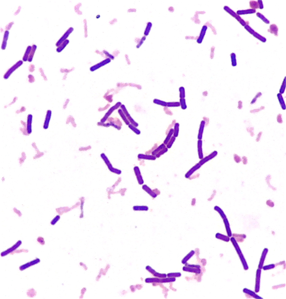
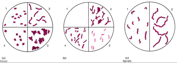
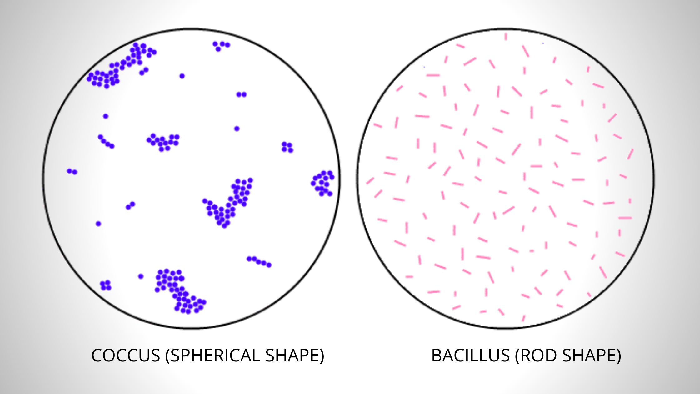
Simple staining is a technique used in microbiology to visualize and differentiate between different types of bacteria. This is accomplished by applying a single stain to the bacterial cells, which can then be observed under a microscope to determine the size, shape, and arrangement of the cells.

There are several different types of stains that can be used for simple staining, including crystal violet, methylene blue, and safranin. These stains are typically applied to a microscope slide containing a sample of the bacterial culture, and then washed off using a decolorizing agent such as ethanol or acetone. The cells that retain the stain appear as a different color under the microscope, allowing researchers to identify and differentiate between different types of bacteria.

One of the main advantages of simple staining is that it is relatively quick and easy to perform, making it a useful tool for identifying and classifying bacterial cultures. It is also relatively inexpensive and can be performed using standard laboratory equipment, such as a microscope and microscope slides.

However, simple staining has some limitations. It is not very specific and cannot distinguish between different types of bacteria with similar morphologies, such as different species within the same genus. In addition, it does not provide any information about the biochemical or metabolic properties of the bacteria. For more detailed analysis, more complex techniques such as gram staining or biochemical testing may be required.

Overall, simple staining is a useful tool for visualizing and differentiating between different types of bacteria, and is an important technique in the field of microbiology. It is frequently used as a first step in the identification and classification of bacterial cultures, and can provide valuable information about the size, shape, and arrangement of bacterial cells.
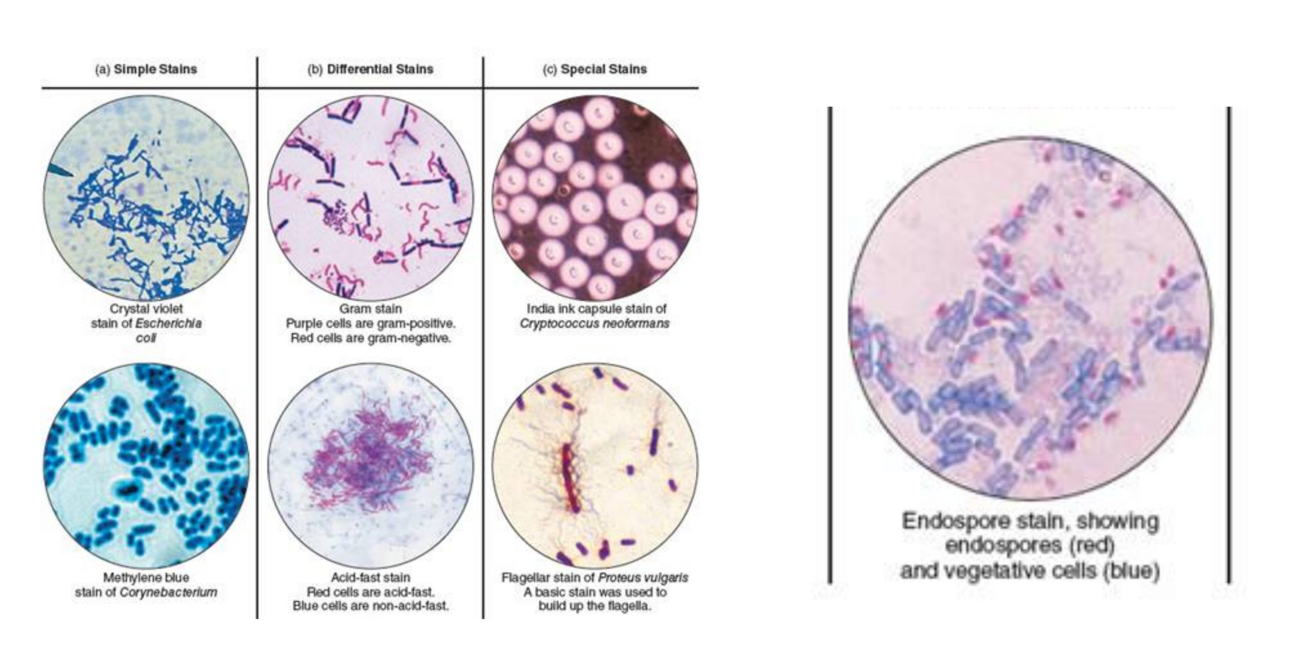
Simple Staining

what is simple staining in microbiology

The Gram stain is a differential stain, as opposed to the simple stain which uses 1 dye. Since the surface of most bacterial cells is negatively charged, these positively charged stains adhere readily to the cell surface. Most of the content available on the recnotes. True to its name, the simple stain is a very simple staining procedure involving a single stain solution. Acid-fast bacillus It distinguishes acid-fast bacteria such as Mycobacterium spp from non-acid fast bacteria; which do not stain well by the Gram staining. Thus acidic conditions favor the work of acidic dye stain whereas alkaline pH works well with basic dyes. II Add immersion oil to the top of the smear when viewing with a 100× immersion lens Stain is too light or too dark I Experiment with incubation time the longer the dye is present the darker the stain No cells are found within the smear I Broth culture was used and not mixed before the sample was taken some cells float on top or sink as a pellet to the bottom of a tube.


Next

What Is Simple Staining Of Bacteria Its Principle, And Procedure?

what is simple staining in microbiology

Simple Staining Most types of cells do not have much natural pigment and are therefore difficult to see under the light microscope unless they are stained. Focus at lowest power and then increase one objective magnification at a time. Alternatively, in simple negative staining, acidic dyes such as nigrosin or congo red are used. Store up to 1 year at room temperature, away from direct sunlight. Acridine orange binds to nucleic acid and stains them.

Next

Different Staining Methods used in Microbiology

what is simple staining in microbiology

CAUTION: Dyes used for bacteriological staining are usually aniline dyes so they are potentially carcinogenic and should be handled carefully. A good stained smear should be somewhat difficult to see with the naked eye on the surface of a microscope slide. Several types of stains can beused to make bacterial cells more visible. Simple vs Differential Staining Simple Staining Differential Staining Definition It is a type of staining that uses only one dye. Examples of simple stains include safranin, methylene blue, crystal violet etc. Acidic stains can stain positively charged molecules in cells including some proteins.

Next

16: Simple Stain

what is simple staining in microbiology

RECOMMENDATION: You may find it helpful to draw a circle wax pencil is best on the opposite side of the slide where you will spread your smear. Be sure the bottom of the slide is clean. If methylene blue is used, some granules in the interior of the cells of some bacteria may appear more deeply stained than the rest of the cell, which is due to the presence of different chemical substances. The simple stain can be used as a quick and easy way to determine the cell shape, size, and arrangement of bacteria. Any basic dye, such as methylene blue, safranin, or crystal violet, can be used to color the bacterial cells. There is an attraction between the positive stain and the negative bacterial cell in simple staining, which results in the observation of coloured bacteria with a bright background. Principle of Simple Staining In simple staining, the bacterial smear is stained with a single reagent, which produces a distinctive contrast between the organism and its background.

Next

Simple staining technique

what is simple staining in microbiology

Tap water is often suggested for the rinsing of stains since it is readily available and the type of water used for rinsing does not affect the outcome of staining results. For more detailed information, consult page 93 of your lab manual. As the simple stains are positively charged, they usually termed as positive or cationic dyes. During this step, hold the slide parallel to the stream of water; in this way you can reduce the loss of organisms from the preparation. Simple staining involves directly staining the bacterial cell with a positively charged dye in order to see bacterial detail, in contrast to negative staining where the bacteria remain unstained against a dark background. What is Simple Staining? Negative stains produce a darker background and cells appear light and unstained.

Next

Simple Stain Flashcards

what is simple staining in microbiology

Identification of these features can lead to a diagnosis and aid in treatment of a patient. For example, when the bacteria retain the safranin colour, they appear pink-red, and the same goes with the other stains. We recycle our microscope slides and the smears have to be removed from the glass. The colour of a stain will decide the colour of a specimen that has to be identified. Simple Stain The simple stain can be used to determine cell shape, size, and arrangement.

Next

Welcome to Microbugz

what is simple staining in microbiology

Be sure to mix a broth culture before taking a sample because many bacteria are not spread evenly throughout the broth, and you may miss your sample. To heat-fix a sample, after a thin layer of the specimen is spread on the slide called a smear or emulsion , and the slide is then briefly heated over a heat source. One needs to be careful about thick smears when taking the specimen from an agar medium. Troubleshooting Common problems that occur throughout the protocol and possible solutions are outlined in Table Problem Solution Cannot bring cells into focus I Make sure the smear side of the slide is on top. It is also used for the detection of mycoplasmas cell wall deficient bacteria.

Next

Difference between Simple and Differential Staining

what is simple staining in microbiology

Dyes bind with cellular constituents producing color contrast and increasing their visibility. It is commonly used to colour most bacteria. The results of simple staining are based on the type of basic stain that has been used. In order to make the slide grease free, wash it, rinse with alcohol and then clean with filter paper to make it dry. The spirilla image does not show flagella.

Next

Simple Staining: Principle, Procedure, Uses, Advantages & Disadvantages

what is simple staining in microbiology

Center the area to be studied, apply immersion oil directly to the smear, and focus the smear under oil with the 100X objective. This step facilitates the fixing of the smear to the slide. Blot the smear slide with your bibulous paper pad. For example, when the bacteria retain the safranin colour, they appear pink-red, and the same goes with the other stains. Methylene blue or malachite green is used as the counterstain. It helps to differentiate between different types of bacterial species.

Next

9: Simple Stain

what is simple staining in microbiology

Staining of Bacteria It is the last and the most crucial step, in which one can identify the morphological characteristics of the bacteria through microscopic examination, once the cells get stained. The results of simple staining are based on the type of basic stain that has been used. Through simple staining, we cannot classify a particular type of organism. This staining either colours the cells or colours the background. In some circumstances e.

Next