Ligament of treves. Sir Frederick Treves, 1st Baronet 2022-12-13
Ligament of treves Rating:
6,8/10
875
reviews
The ligament of Treves, also known as the mesentericoparietal ligament, is a small but important structure in the human body. It is located in the abdomen and helps to support and stabilize the small intestine.
The small intestine is a long, slender tube that is responsible for the absorption of nutrients from the food we eat. It is located within the abdominal cavity, and is supported and held in place by a series of ligaments, muscles, and connective tissue. The ligament of Treves is one of these structures, and it plays a crucial role in keeping the small intestine in place.
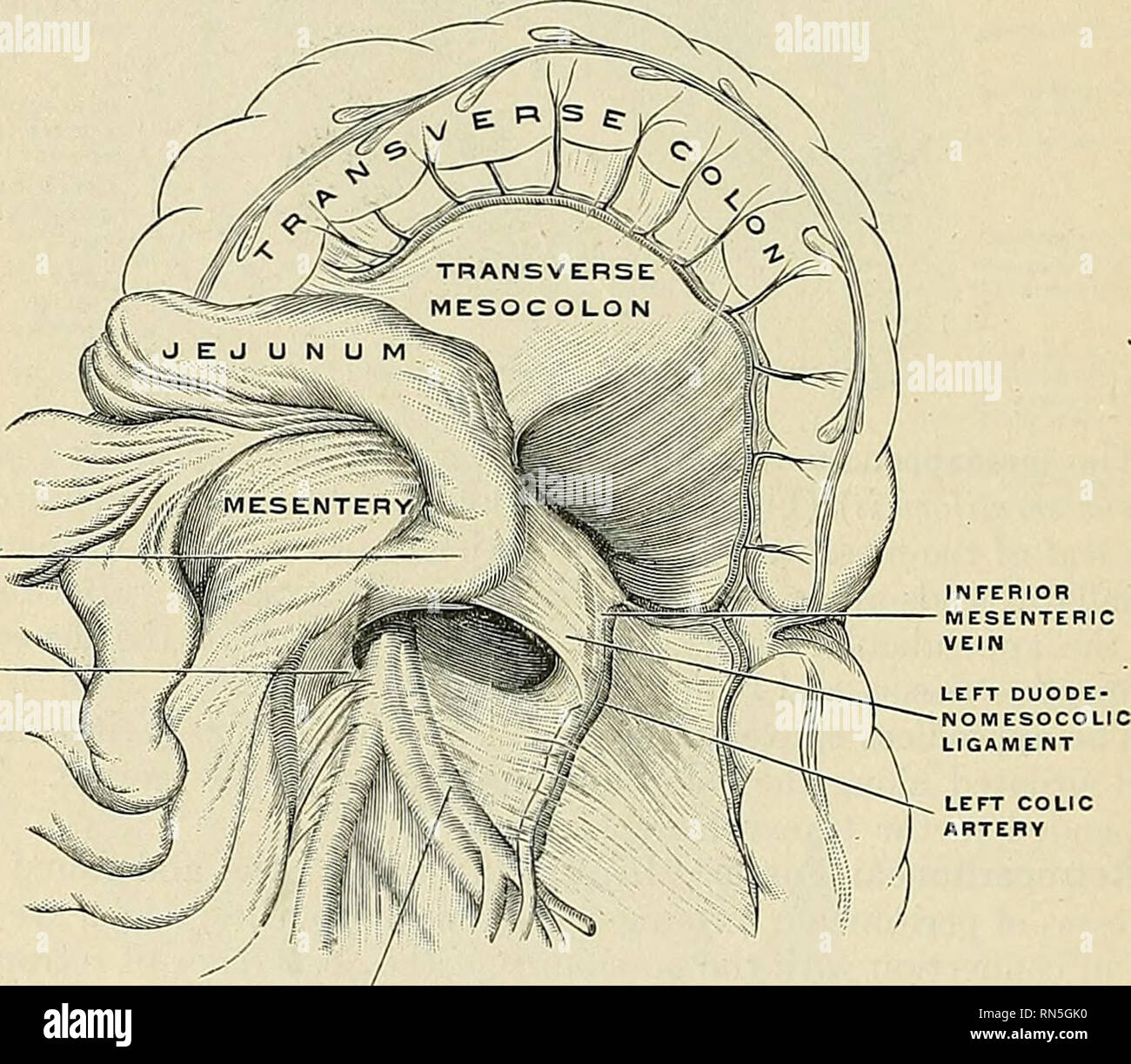
The ligament of Treves is a thin, fibrous band that runs from the mesentery (a sheet of connective tissue that attaches the small intestine to the abdominal wall) to the posterior abdominal wall. It is located in the lower left quadrant of the abdomen, just below the left kidney.
The ligament of Treves is important because it helps to prevent the small intestine from becoming displaced or twisted, which can cause serious problems. It also helps to support the small intestine and keep it in place during movement and other physical activities.
In addition to its structural function, the ligament of Treves also plays a role in the circulation of blood and lymph to the small intestine. It contains small blood vessels and lymphatic vessels that help to nourish and support the small intestine.
Despite its small size and relatively simple structure, the ligament of Treves is an important part of the human body. It helps to support and stabilize the small intestine, and is essential for proper digestion and nutrient absorption.
Ligament
Journal Of Emergencies, Trauma, And Shock, 4 3 , 440. It forms the posterior border of the femoral canal. Its composite bands extend between the adjoining fibular and tibial surfaces. The support it receives comes in two flavours: dynamic and passive. The ligament is made up of two bands that originate from the transverse processes of L5. Ligamentum venosum Synonyms: Ligament of Arantius, Venous ligament of liver , In utero, the ductus venosum was responsible for shunting blood from the left portal vein to the left hepatic vein, thereby bypassing the hepatic circulation. Some anatomists were only able to identify smooth muscle fibers.
The uterus is only one of many organs in the reproductive system. This phenomenon is characterized by free peritoneal air outlining the falciform ligament. It can often find damage or disease in bones and a surrounding ligament, tendon, or muscle. While this is a relatively small structure, it has clinical implications in surgical procedures and in rare cases of small bowel obstruction. The lower part of the muscle may be constant with the rounded as well as the longitudinal layer of muscle lying towards the duodenojejunal junction.
Abdom Imaging, 32 1 , 59-65. The named ligaments involved in stabilizing this joint are the interosseous, medial, lateral and cervical ligaments. Â Distally, the triangular ligament integrates with the iliofemoral ligament and occasionally the neck of the femur. It is formed from the left halves of the two layers of the coronary ligament. This structure is a reflection of the diaphragmatic peritoneum that attaches to the superior and posterior aspects of the right lobe of the liver. Any abnormal deviation from the norm has a high potential of a pathological development that will severely impact the health and well being of the female. Journal Of Emergencies, Trauma, And Shock, 4 3 , 440.
The tibiocalcaneal also known as the intermediate part takes a vertical route to insert along the upper border of sustentaculum tali a bony projection of the calcaneus. Wenzel Treitz an Austrian physician. Clinically oriented anatomy 5th ed. What are the symptoms of a collateral ligament injury? It has attachments to the roughened posterior aspect of the patella, the distal apex of the patella as well as its margins. It is also known as the Silver sign.
Ligaments of the uterus: Function and clinical cases
The only difference among them is in the connection that they make internally: ligaments connect one bone to another bone, where the tendons connect muscle to bone, and fasciae connect muscles to other muscles. The ligaments of the uterus have an important role in both. In some cases, the ligamentum teres hepatis fails to degenerate and remains patent. The ligament of Treitz the suspensory ligament of the Duodenum : anatomic and radiographic correlation. Unusual positions of the uterus increase the chance of a prolapse because the bladder cannot support it during an episode of increasing pressure inside the abdominal cavity. The pubocervical, transverse cervical and uterosacral ligaments radiate outwards from the cervix towards the pelvic side walls, like the spokes of a wheel. Ligaments, whether they connect bones or organs, help the body maintain stability.
Quantitative Anatomic Analysis of the Native Ligamentum Teres. The two leaves unite to form the falciform ligament, which descends on the anterior surface of the liver between the two lobes. Superiorly, the falciform ligament is attached to the visceral aspect of the anterior abdominal wall just an inch to the right of the median plane and the inferior surface of the diaphragm. Learn the anatomy of the liver and entire digestive system in an easy and effective by using Kenhub's On the right lobe of the liver, the anterior and posterior layers of the coronary ligament of the liver join to form the right triangular ligament. Gray's Anatomy: The Anatomical Basis of Clinical Practice, 41st edition.
Ligament of Treitz: (Detailed Information Read Here)
It finally ends in the duodenojejunal flexure and the distal duodenum. The support it receives comes in two flavours: dynamic and passive. Ligaments are similar to fasciae and tendons as they are all made of connective tissue. Hence, it is virtually impossible to image 2,3. This is probably why the patellar ligament is often incorrectly referred to as a tendon. The attachment of the coronal ligament to the liver and its division into the two layers creates a triangular area that is devoid of peritoneum.
Histologically, the suspensory muscle of the duodenum is comprised of With around 10 000 anatomy terms, you probably think it's barely possible to memorize them! Tibiofibular ligaments There are two short ligaments that attach the medial part of the fibular head to the adjacent region of the tibial head known as the superior tibiofibular ligaments ligaments of the fibular head. These are the pubocervical, transverse cervical and uterosacral ligaments. The developing human 9th ed. How well do you know the surrounding structures? Histological assessment of the band reveals that Hilfsmuskel is made up of The second part of the ligament of Treitz is a thin muscular band originating from the celiac trunk as a connective tissue band. Frederick Arthur Crisp, 1905, p. It is actually this second part of the structure that is responsible for suspending the duodenojejunal flexure. Frequently Asked Questions When should I call a healthcare provider? Morphology of the ligament of Treitz likely depends on its fetal topographical relationship with the left adrenal gland and liver caudate lobe as well as the developing lymphatic tissues: a histological study using human fetuses.