Parietal branches of abdominal aorta. The Abdominal Aorta: Function And Anatomy 2022-12-31
Parietal branches of abdominal aorta Rating:
5,5/10
1899
reviews
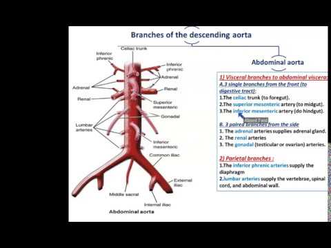
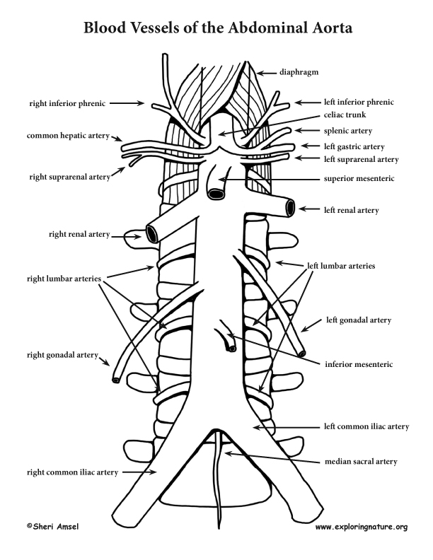
The abdominal aorta is the main blood vessel that carries oxygenated blood from the heart to the lower half of the body. It is a large and important vessel that branches off into smaller arteries, which supply blood to the various organs and tissues of the abdomen. One of these branches is called the parietal branch, which is a smaller artery that supplies blood to the abdominal wall.
The parietal branches of the abdominal aorta arise from the main vessel near the level of the umbilicus, or belly button. These branches run laterally along the abdominal wall, supplying blood to the muscles and skin of the abdominal wall. They also supply blood to the peritoneum, which is the thin layer of tissue that lines the inside of the abdomen and covers most of the abdominal organs.
There are several different parietal branches of the abdominal aorta, including the inferior epigastric artery and the superior epigastric artery. The inferior epigastric artery runs downward along the inner aspect of the abdominal wall, while the superior epigastric artery runs upward along the outer aspect of the abdominal wall. Both of these arteries supply blood to the muscles and skin of the abdominal wall, as well as to the peritoneum.
The parietal branches of the abdominal aorta are important for maintaining the health and function of the abdominal wall and its associated organs and tissues. They provide a vital source of oxygen and nutrients, which are necessary for the proper functioning of these structures. In addition, the parietal branches of the abdominal aorta are important for maintaining blood pressure and blood flow within the abdomen.
In summary, the parietal branches of the abdominal aorta are small arteries that branch off from the main abdominal aorta and supply blood to the muscles and skin of the abdominal wall, as well as to the peritoneum. These branches are important for maintaining the health and function of the abdominal wall and its associated organs and tissues, and are vital for maintaining proper blood flow and blood pressure within the abdomen.
The Three Paired Visceral Branches Of The Abdominal Aorta
Each vessel gives off some small inferior suprarenal branches to the suprarenal gland, the ureter, and the surrounding cellular tissue and muscles. Branches The left and right aortic sinuses are dilations in the ascending aorta, located at the level of the aortic valve. The aortic arch is divided into five branches: the right common carotid artery, the left common carotid artery, the left thyrocervical trunk, the left subclavian artery, and the right subclavian artery. It receives the cardiac output from the left ventricle and supplies the body with oxygenated blood via the systemic circulation. The 9 pairs of intercostal arteries supply the intercostal spaces, with the exception of the first and second they are supplied by a branch from the subclavian artery. Immuneglobulin G4-related diseases, according to reports, are one of the most common causes of The deceleration of a seat belt can cause abdominal aortic injury.
The Abdominal Aorta: A Large Blood Vessel That Supplies Blood To The Abdomen Pelvis And Legs
The pulmonary artery carries oxygen-poor blood from the heart to the lungs. Below, it is in relation to the upper border of the pancreas, and the lienal vein. One of these, larger than the rest, is sometimes given off near the tail of the pancreas; it runs from left to right near the posterior surface of the gland, following the course of the pancreatic duct, and is called the arteria pancreatica magna. The abdominal aortic aneurysm AAA is a bulge or swelling in the aortic, the main blood vessel that runs from the heart to the stomach and chest. Unpaired branches: the coeliac, superior mesenteric, inferior mesenteric, and median sacral arteries.
They pass from left to right, between the layers of the gastrolienal ligament, and are distributed to the greater curvature of the stomach, anastomosing with branches of the left gastric and left gastroepiploic arteries. In the fetus these arteries are of large size. The In certain high-risk patients, routine ambulatory visits should include physical exams and According to a systematic review and meta-analysis of over 50 studies evaluating cardiovascular risk associated with abdominal aortic calcification AAC , this condition has been linked to a 20% increase in cardiovascular event mortality. The Important Arteries Of The Abdominal Wall The parietal branches supply essential muscles in the abdominal wall and are located in the abdominal aorta. I received my Ph. The first and often largest visceral branch of the abdominal aorta is the celiac trunk.
What Are The 3 Major Branches Of The Abdominal Aorta?
The end section of the left gastric-glandular artery in the large curvature of the stomach anastomoses with the right gastro-omental artery. Because of the fact that it contains fewer elastin fibers in its wall, the abdominal aorta is the most common type of aortic dissection. Since the normal diameter of the ascending aorta amounts to about 3. Aortic aneurysms are a common cause of death in the left common carotid artery, which is one of the arteries that carry blood to the brain. If you have any symptoms, such as chest pain, shortness of breath, or weakness, see your doctor as soon as possible. Clinical Relevance: Coarctation of the Aorta Coarctation of the aorta refers to narrowing of the vessel, usually at the insertion of the ligamentum arteriosum former ductus arteriosus.
The small intestine and the large intestine begin at the same location in the body, the cecum. In between L1 and L2 Visceral yes post. If left untreated, a large aneurysm can rupture. The most common type of aortic aneurysm is the abdominal aortic aneurysm. The abdominal aorta is the largest blood vessel in the abdomen, and it is the most common type of blood vessel in the abdomen.
The celiac trunk is a branch of the digestive system that functions as a protective branch, as these critical organs cannot function without adequate blood flow. Abdominal Aorta And Its Branches: Functions And Importance The inferior phrenic, sacral, and median branches of the abdominal aorta are all fracture-supporting structures in the abdominal wall. The only branches of the ascending aorta are the coronary arteries, which arise just where it starts. The aortas are separated from the common iliac arteries by an incision in the right and left sides, and a needle is inserted into the right and left sides to be tested. The pancreatic branches rami pancreatici are numerous small vessels derived from the lienal as it runs behind the upper border of the pancreas, supplying its body and tail. The celiac trunk is a short vessel formed by the aorta that passes beneath the median arcuate ligament, just as the aorta enters the abdomen from above at the T12 vertebra level.
Shortly after its origin, the innominate artery divides into the right subclavian and right common carotid arteries. What are the three branches of the aortic arch quizlet? Mesenterica inferior starts from the left semicircle of the abdominal part of the aorta at level III of the lumbar vertebra, goes behind the peritoneum downward and to the left and gives a series of branches to the sigmoid, descending colon and left side of the transverse colon. The largest artery in the body, it is made up of three parts, each with its own distinct shape and direction. Each artery gives off branches to the adrenal gland superior suprarenal artery. It carries oxygen-rich blood from the heart to the rest of the body.
The number of paired branches of the abdominal part of the aorta includes the middle adrenal, renal, and ovarian ovarian arteries. An anterior cerebral artery ACA serves as a unique vessel from a phylogenetic standpoint. It transports blood to the brain and other organs in the head in a variety of ways. The right gastric artery a. It is located in the left pulmonary hilum, pericardium that surrounds the left atrium and esophagus, and is adjacent to the left pulmonary hilum. The celiac trunk is the largest visceral branch of the abdominal aorta and the first to emerge from the abdominal aorta.
What are the 3 major branches of the abdominal aorta?
Patients who have ruptured AAAs have a 5-year survival rate of 97%. At this level, the aorta terminates by bifurcating into the right and left common iliac arteries that supply the lower body. The aortic arch is divided into three branches, one of which is the brachiocephalic trunk, another is the left common carotid artery, and the third is the left subclavian artery. One of the two major arteries that supply blood to the intestines is the inferior mesenteric artery. It is possible that the diameter of the fetal pulmonary arteries will predict the outcome of a congenital diaphragmatic hernia. They give rise to the left and right coronary arteriesthat supply the myocardium. It enters the abdomen through the aortic opening of the diaphragm, which is located beneath the median arcuate ligament between the crura of the diaphragm at T12.
Can you navigate your way through our quiz about the cardiovascular system? The origin and course of the first part of each artery are the same as those of the internal spermatic, but on arriving at the upper opening of the lesser pelvis the ovarian artery passes inward, between the two layers of the ovariopelvic ligament and of the broad ligament of the uterus, to be distributed to the ovary. These branches are obtained from the inferior phrenic, lumbar, and median sacral arteries. A aortic aneurysm is a localized enlargement of the aortic wall that can be fatal. Human ACA formation is more likely the variant of extensive fusion rather than the incidental appearance of primitive forms. Abdominal aorta Synonyms: Abdominal part of aorta, Pars abdominalis aortae The abdominal aorta emerges from the aortic orifice of the diaphragm at the level of T12 vertebra. Lumbar Arteries: Paired arteries of usually 4—5 segments which supply the abdominal wall.