Sds page experiment report. SDS 2022-12-09

Sds page experiment report Rating: 6,8/10 1545 reviews

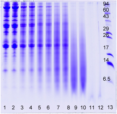
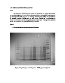
SDS PAGE (sodium dodecyl sulfate polyacrylamide gel electrophoresis) is a commonly used technique in molecular biology laboratories for separating and analyzing proteins based on their size and charge. In this experiment, a protein sample is mixed with SDS, a detergent that coats the protein and gives it a uniform negative charge. The protein-SDS mixture is then loaded onto a polyacrylamide gel, which is placed in a special apparatus and subjected to an electric current. As the protein moves through the gel, it separates into individual bands based on its size, with the smaller proteins moving faster through the gel than the larger ones.

To begin the experiment, the necessary materials and equipment are assembled, including a protein sample, SDS, polyacrylamide gel, a gel apparatus, and a power supply. The protein sample is then mixed with SDS according to the manufacturer's instructions, taking care to ensure that the SDS is evenly distributed throughout the sample.

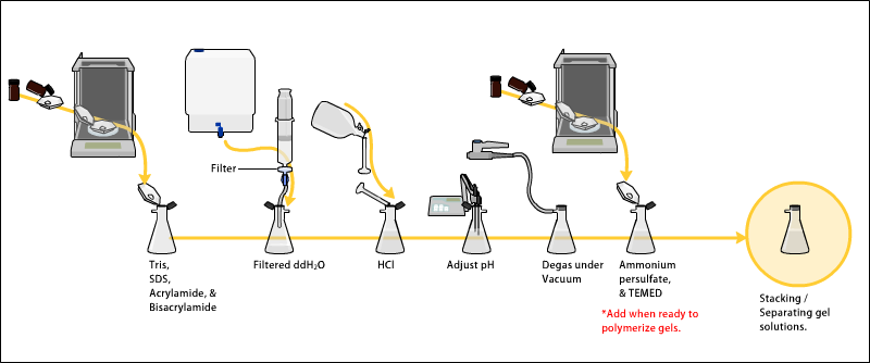
Next, the polyacrylamide gel is prepared according to the manufacturer's instructions, taking care to ensure that the gel is properly formed and free of defects. Once the gel is prepared, it is placed in the gel apparatus and the protein-SDS mixture is carefully loaded onto the gel. The apparatus is then connected to the power supply, and the electric current is turned on.

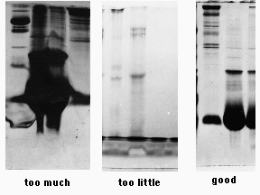
As the protein moves through the gel, it separates into individual bands based on its size. These bands can be visualized using a special stain or by placing the gel under UV light, which causes the protein to fluoresce.

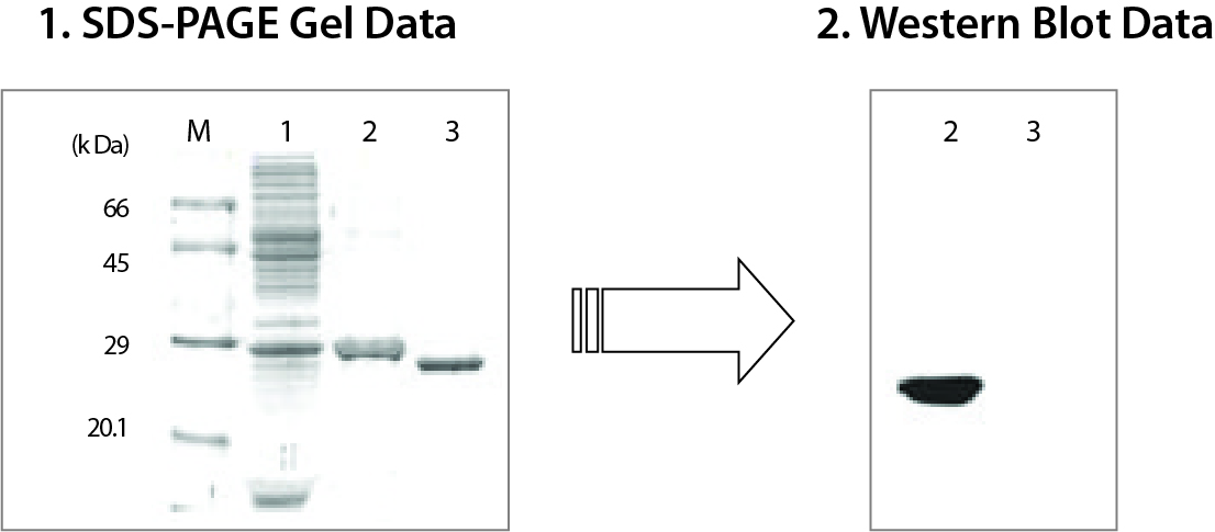
The results of the SDS PAGE experiment can be analyzed and interpreted in a number of ways. The size of the protein bands can be measured and compared to known standards, allowing researchers to determine the size of the proteins in the sample. In addition, the presence or absence of certain protein bands can provide information about the purity of the sample, as well as any post-translational modifications that may have occurred.

Overall, SDS PAGE is a powerful tool for separating and analyzing proteins, and is an important technique in many areas of molecular biology research. It is a reliable and reproducible method that allows researchers to gain valuable insights into the structure and function of proteins, which is critical for understanding biological processes at the molecular level.

Methods for SDS

sds page experiment report

A protein first runs through the stacking gel, where the samples spread out. Also to understand the assessment of the protein size and purity. The running buffer surrounds the gel during electrophoresis. The blue bands seen are the protein bands that have been transferred. This technique uses a discontinuous gel consisting of a resolving gel and a separating gel, both of which had to be made prior to the start of the experiment. .

Next

SDS

sds page experiment report

Find Out How UKEssays. Creating Ideal Separation Conditions: Some Factors to Consider Given its importance in determining the molecular weight of an unknown protein sample, you need to select the appropriate separation conditions when doing your experiment. Question: How to Determine Molecular Weight of Protein with SDS-Page? Neal Burnette April 1981. Discussion In this experiment, it was concluded that the unknown sample was Î’- Galactosidase. Table 2: Unknown Protein Sample. Place the gel in Coomassie Brilliant Blue G-250 staining solution in a plastic container and rock for several hours or follow Advice below.

Next

Polyacrylamide Gel Electrophoresis: Protein Separation

sds page experiment report

Polyacrylamide Gel Electrophoresis Polyacrylamide gel electrophoresis PAGE is probably the most common analytical technique used to separate and characterize proteins. The known protein standards are identified and act as markers for the unknown sample. Polyacrylamide Gel Electrophoresis SDS-PAGE October 8, 2018 BIO 3522 Section Fall 2018 Introduction: The purpose of this lab is to learn how to purify a unknown protein sample, and to learn how to find the molecular weight of a protein. Lanes 4-12 show protein expression results from the plasmids indicated in the absence or presence of para-bromophenylalanine pBrF. The cassette was then placed into the tank with the grey panel at the cathode side of the tank, which is then filled with transfer buffer. Electrophoresis is the process of separating proteins by forcing them to move through a polymeric gel where they move per the electrical charge of the protein and shape, or size.

Next

SDS

sds page experiment report

The Protein Man Says: How can you estimate the molecular weight of an unknown protein? Air bubbles were removed before and after a further piece of saturated filter paper was placed on top of the nitrocellulose. Butanol was poured on top of the separating gel and it was allowed to polymerise. This gel was then allowed to polymerise for 45 minutes and then the comb was removed, and washed with distilled water once more. This is a useful technique for biochemists, because it allows them to purify a protein from a sample solution using the sample buffer in the gel. Cleavage of Structural Proteins during the Assembly of the Head of Bacteriophage T4. Consequently, the rate at which the SDS coated proteins migrate in the gel is purely dependent on size. How do you know that there is only one variable in this separation? Protein standards measured the following distances: Myosin — 0.

Next

SDS PAGE lab report

sds page experiment report

The graph shows the results of the SDS-PAGE. From the samples that provided were the distinct bands made on the gel at various distances. What is the variable this time? The six standard proteinswere identified as: Myosin 200 kDa , a motor protein found in eukaryotic tissues, and plays a key role in muscle contraction in the striated and smooth muscle cells along with actin; Î’-Galactosidase 116 kDa a hydrolase enzyme, which catalyses the hydrolysis of Î’-galactosides into monosaccharides and an essential human body enzyme which is also produced in E. By the end of this lab, we will be able to use the data that we collect throughout the experiment to calculate the molecular weight of the protein sample and graph the Log of MW vs the Rf values found in the experiment. Western Blot, also known as Immunoblot, is a biochemical technique which targets a specific protein which has been the result of a separation technique such as SDS-PAGE. The gels are usually prepared with the top portion of the gel under the sample wells made less dense than the remainder of the gel below that is intentionally made denser.

Next

Determining Protein Molecular Weight with SDS

sds page experiment report

The solution was then poured to within 1cm of the comb. Describe functions of each constituent. Introduction Gel electrophoresis is a separation technique used by biologist to separate proteins according to the size of the unknown proteins. As they move through the resolving gel they separate by size. Because the proteins are not denatured, they are less predictable in the way they move through the gel, and take much longer.


Next

SDS PAGE LAB blog.sigma-systems.com

sds page experiment report

Last but not least, the electrophoresis would be applied to cause the proteins to migrate across the gel. The resolving lower gel is where the proteins migrate to and separate into different sizes. Then add 50 µL of 2X SDS dye. Gel filtration chromatography and ultracentrifugation are previous experiments that also carry out separation by size, however, this experiment is used to determine more accurate differences in molecular weight. The gel was overlain butanol and left to polymerise. The bisacrylamide introduces crosslinks between polyacrylamide chains. After the gel has been prepared as outlined in the Instruction Protocol, 20 µL of the supernatant can be placed in each well of the polyacrylamide gel.


Next

1.15: SDS

sds page experiment report

Based on the values obtained for the bands in the standard, the logarithm of the molecular weight of an SDS-denatured polypeptide and its relative migration distance Rf is plotted into a graph. The quantity of acrylamide in the resolving gel defines how well the separation takes place since higher concentrations of acrylamide restrict the mobility of proteins. Results SDS-PAGE Following SDS-PAGE, and the removal of the gel, protein standards were identified and the known Molecular Weights , measured in kilo Daltons kDa , were calculated and given to the class. Alternatively, an appropriate software may also be used to determine the Rf values of the resulting bands. The SDS denatures the secondary and tertiary structure of the protein, excluding disulphide bridges, and binds at a ratio of 1. This table shows the unknown proteins, A and B which were present in the unknown sample.


Next

Polyacrylamide Gel Electrophoresis (SDS

sds page experiment report

To run the gel, follow the Bio-Rad Protean II Instruction Protocol. Identifying Proteins The blotted membrane was placed in the staining bath and covered with blocking solution for 10 minutes. The membrane is then treated with specific antibody solution which is known to target the protein in question. Here's everything you need to know. SDS-PAGE was run at 200V for approx.

Next

sds page experiment report

Unknown Proteins Molecular Weight MW D Log10 MW Distance Migrated cm Protein A 63,100 4. SDS-PAGE Sodium dodecyl sulfate SDS is an amphipathic detergent. A final piece of saturated blotting paper was layered on top, again, avoiding air bubbles. From simple essay plans, through to full dissertations, you can guarantee we have a service perfectly matched to your needs. These include colorimetric detection which uses an enzyme such as peroxidase to stain the membrane and radioactive detection, which applies x-rays to the membrane and colours the target protein s a dark colour.

Next