Structure of eye and ear. A&P/2 LAB 1: Anatomy of the Eye & Ear Flashcards 2022-12-28

Structure of eye and ear Rating: 7,3/10 1067 reviews

The eye and the ear are two of the most important sensory organs in the human body. They allow us to perceive and interpret the world around us through sight and sound. While both the eye and the ear serve different functions, they share some similar structural features. In this essay, we will discuss the structure of the eye and the ear in detail.

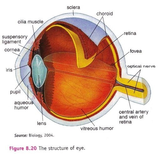
The eye is a complex organ that is responsible for capturing and processing light stimuli. It is made up of several different parts, including the cornea, the iris, the pupil, the lens, the retina, and the optic nerve.

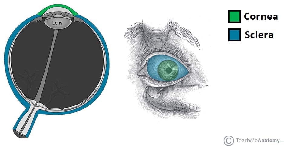
The cornea is the clear outer layer of the eye that helps to protect it and also helps to focus light as it enters the eye. The iris is the colored part of the eye that is responsible for controlling the size of the pupil, which is the small, circular opening in the center of the iris. The pupil adjusts the amount of light that enters the eye by contracting and dilating in response to changes in light intensity.

The lens is a transparent, curved structure that is located behind the pupil and is responsible for focusing light onto the retina. The retina is a thin layer of tissue at the back of the eye that contains photoreceptor cells called rods and cones. These cells are responsible for converting light into electrical signals that are sent to the brain via the optic nerve.

The ear is another important sensory organ that is responsible for the perception of sound. It is made up of three main parts: the outer ear, the middle ear, and the inner ear.

The outer ear consists of the pinna (the visible part of the ear) and the ear canal. The pinna is the external part of the ear that is responsible for collecting and directing sound waves into the ear canal. The ear canal is a short, narrow tube that leads from the outer ear to the middle ear.

The middle ear is a small, air-filled chamber that is located behind the eardrum. It contains three tiny bones called the ossicles (the malleus, incus, and stapes). These bones amplify the sound waves that are transmitted through the ear canal and eardrum, and transmit them to the inner ear.

The inner ear is the most complex and delicate part of the ear. It is made up of the cochlea, which is responsible for converting sound waves into electrical signals, and the vestibular system, which is responsible for maintaining balance and detecting the body's position in space.

In conclusion, the eye and the ear are two important sensory organs that allow us to perceive and interpret the world around us. While they serve different functions, they share some structural similarities and are both essential for our daily lives.

Elementary structure and functions of eye and ear

structure of eye and ear

As seen in the eye, it is the white part. In the center, we have a vascular tunic, or we can reach it uvea, and it is brief of the choroid, ciliary body and in this iris is also there. In addition to covering the white of the eye, the conjunctiva is a thin membrane that traces the edges of the eyelids and covers the corneal epithelium. . By pulling on the medial rectus muscle, for example, you can look at your nose while pulling your eyeball to the side. Glutamate, a neurotransmitter, is released due to Ca ++ ion movement inside the cell.

Next

Structure of the eye and ear Free Essays

structure of eye and ear

They are hearing and balancing. Cataracts are very common in older adults. Most of the eye is a mechanical device that is Premium Auditory system Visual perception Brain Head, Ears, Eyes, Nose, Throat Physical Assessment surgery. Video used with permission. It has few more parts like the oval window, incus, etc.

Next

8.2 Eye and Ear Basic Concepts

structure of eye and ear

Smoking has been a common habit in the modern world. The inner layer is called the retina, which are the cells sensitive to light rods and cones , covered by an outer sheet of cuboidal epithelial cells containing melanin. Ototoxic medicationscan also cause sensorineural hearing loss by affecting the hair cells in the cochlea. Please review all the terms from the lab guide and pay special attention to the key terms that carry over from our previous lab sessions. Â Acute otitis media typically occurs after an upper respiratory infection when the Eustachian tube becomes inflamed and the middle ear fills with fluid, causing ear pain and irritability. In the anterior wall the eustachian tube opening can be found, connecting the middle ear to the nasopharynx.

Next

Anatomy of Eye and Ear

structure of eye and ear

Which structure of the ear is responsible for the balance? Also, stapes is the smallest bone in the entire human body. Vision begins when light is reflected off a surface and enters the eye through the cornea, refracting the rays through the pupil. Conclusion In the above chapter, we have studied the Elementary structure and function of the eye and ear examples. Some of these images show a superior or lateral view of the orbit, while others show cross-sectional images of the eye. The middle ear has three tiny bones, and steps are one among them. Your eyes can help you find your best friend at a crowded concert.

Next

Structure Of The Eye And Ear, Sample of Essays

structure of eye and ear

In the cornea, near the anterior chamber, a trabecular meshwork drains aqueous humor continuously. The idea is supported by the case of SB. There are six extrinsic eye muscles to help move each Eye; the superior rectus, inferior rectus, lateral rectus, medial rectus, superior oblique and inferior oblique. This occlusion often causes symptoms such as hearing loss, ear fullness, and itching. The three main structures of the ear are the outer ear, middle ear and inner ear. Treatment includes over-the-counter and prescription eye drops, as well as lifestyle changes to decrease the dryness of the eyes.


Next

Structure And Function Of Eye And Ear

structure of eye and ear

Besides being yellow, it is also highly pigmented. The eyelashes help protect from foreign objects and perspiration, as well do the Eyebrows. Cornea, aqueous humor and biconvex lens completely refract the light rays coming from the object. This is one of the most important topics from the examination point of view. Auditory systems involved in sound wave that transduced by drum ear into some kind of vibration that eventually gets converted back into wave what we perceive as noise.

Next

Human Ear Structure and Function

structure of eye and ear

These muscles function to move the eye so that we can direct our visual attention. It connects to the surface of the eyeball and these six extrinsic eye muscles. During myringotomy surgery, a tympanostomy tube is placed in the tympanic membrane to drain fluid from the middle ear and prevent infection from developing. Structure of the eye are comprehended in three covers or coats within with it consists of further three transparent coats. These are all on the wall of the eyeball, the fibrous tunic consist of the cornea and the sclera. The four preceding articles all discuss the obligations of individuals within a society. Its main function is to amplify and transmit the sound waves to the internal ear.

Next

Structure of the eye and ear

structure of eye and ear

Lens There is a lens situated between the vitreous humor and the pupil in the anterior portion of the eye. As the ear is a sensitive organ in our body, it plays a vital role. The middle layer encompasses the blood-supplying choroid and pigmented iris. The Lacrimal apparatus is a group of small structures, when humans Express emotions with tears, the Lacrimal apparatus helps to produce and then Drain those tears away. Symptoms of glaucoma include gradual loss of peripheral vision. A: small ducts bring tears to the front and blinking help spread the tears over the surface Q: what enzyme inhibits the growth of bacteria on the surface of the eyes? This is a common view of both of the theories of perception. Acute Otitis Media Acute otitis media is the medical diagnosis for an middle ear infection.

Next

Structure and Functions of Eye & Ear and their Disorders : Pharmaguideline

structure of eye and ear

Because of this, crying can make the nose run. It is quite necessary to study the structure and function of the human eye and ear. Structure of Eye The Within these coats lie the flexible lens, aqueous humor and the vitreous body. However, the treatment goals are to control pain and treat infection with antibiotics if a bacterial infection is present. A depression known as the fovea centralis is located in the macula, which contains concentrated light-detecting cells. EAR INFECTION: An ear infection is a common type of infection and is the result of an invasion of bacteria or viruses into the ear.


Next